Chest CT

Understand your chest CT — in language that actually makes sense

Upload your chest CT (the folder of DICOM slices) and get a clear, plain-language explanation of what the findings mean: lung nodules, ground-glass, emphysema, scarring, and the terms that sound worse than they are.

GDPR compliant Encrypted Delete anytime
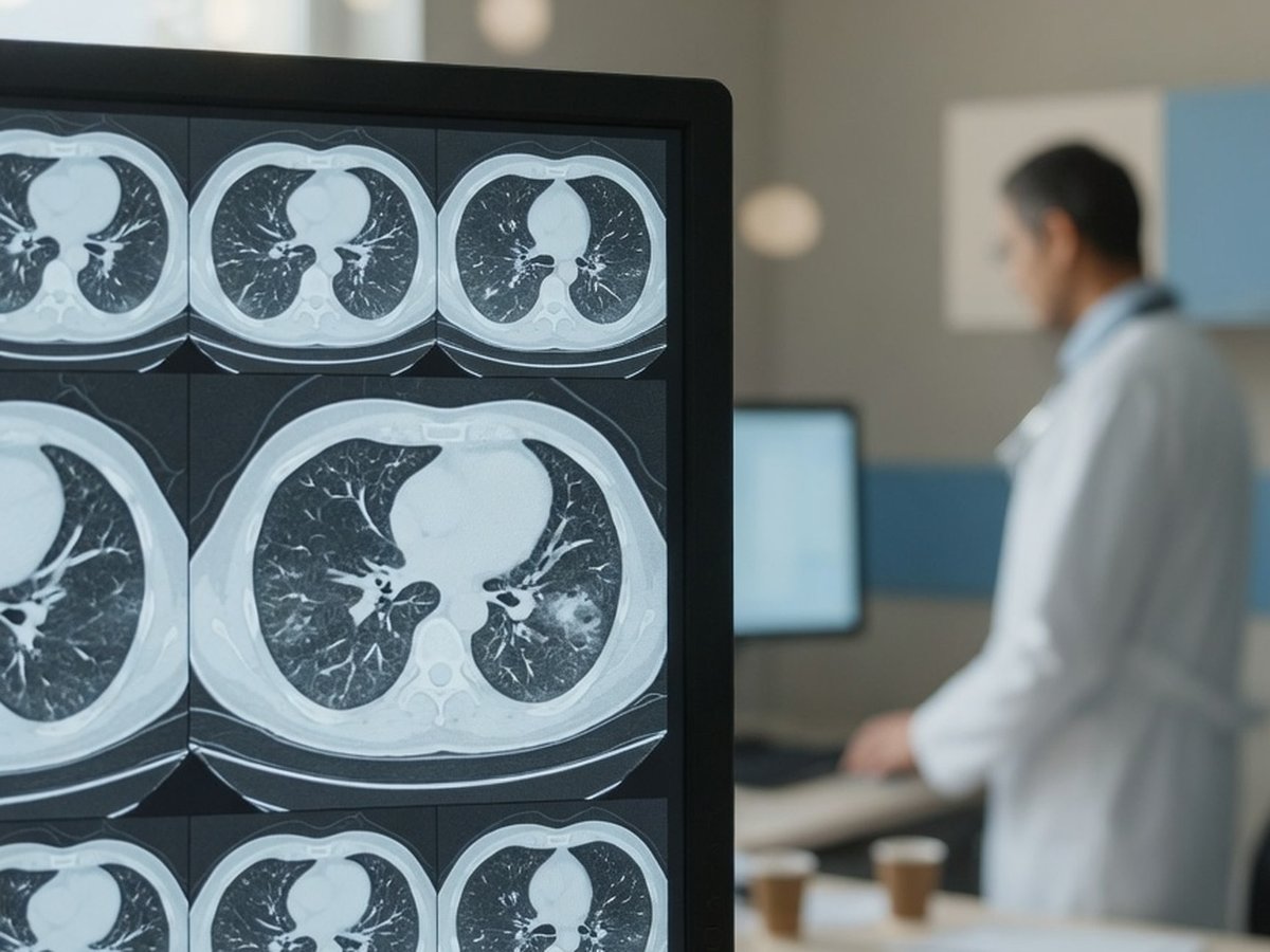
Chest CT cross-sections on a radiology reading-room monitor — ReadYourLab AI chest CT interpretation

Why chest CT reports are so hard to read

A chest CT captures hundreds of thin cross-sectional slices, so it sees far more than a plain X-ray — and its report is a dense, structure-by-structure list: "a 5 mm solid nodule in the right upper lobe, mild centrilobular emphysema, no pleural effusion, unremarkable mediastinum."

Because CT is so sensitive, it turns up small things constantly — a tiny nodule, a fleck of old scar, a bit of ground-glass — and most of these are benign or incidental. The report rarely tells you, in plain words, which findings are trivial and which deserve a follow-up.

ReadYourLab reads your slices with medical AI and rewrites the report in plain language: what each finding is, how common it is, and which ones are worth a prompt conversation with your doctor. It's an informed first look to help you prepare — not a diagnosis.

Common chest CT findings — explained

The terms that show up most often on chest CT reports, and what they usually mean.

Pulmonary nodule

Usually followed up

A small round spot in the lung. On CT they're found very often, and the large majority are benign — old scars or healed infection. Because a minority are not, they're commonly followed with a repeat CT to check for change over time; size guides how closely.

Full explanation

Ground-glass opacity

Context matters

A hazy area that doesn't fully block the lung markings behind it. Very non-specific — it can reflect infection, inflammation, or scarring, and is often just followed up. What it means depends heavily on the surrounding findings and your symptoms.

Full explanation

Emphysema

Chronic finding

Areas where lung tissue has been destroyed and looks like small dark holes, typically from smoking. A chronic, gradual change rather than an acute problem, and part of the COPD picture.

Full explanation

Calcified granuloma

Benign scar

A small, hard, calcium-containing spot — the healed scar of an old, long-resolved infection. Extremely common, completely benign, and needs no action; it's essentially the lung's version of a scar.

Full explanation

Consolidation

Often infection

An area of lung filled with fluid, pus, or cells instead of air, so it looks solid white. The classic appearance of pneumonia; usually treated and then re-imaged to confirm it clears.

Full explanation

Bronchiectasis

Chronic finding

Airways that have become permanently widened and thickened, often after repeated infections. A chronic condition managed over time rather than an emergency.

Full explanation

Pleural effusion

Cause matters

Fluid between the lung and chest wall. Small amounts are common after infection or surgery; larger ones prompt a search for the cause. It's the underlying reason, not the fluid itself, that guides treatment.

Full explanation

Lymphadenopathy

Read in context

Lymph nodes that look larger than usual. Frequently a reaction to infection or inflammation and often benign, but sometimes followed up depending on size, pattern, and the rest of the scan.

Full explanation

Descriptions here are general education, not a reading of your specific CT. Always review your report with your doctor, and seek urgent care for severe breathlessness, chest pain, or coughing up blood.

How lung nodules are followed

Chest CT doesn't use a single numbered severity scale, but nodules are followed by well-known size-based rules.

Nodule size & Fleischner-style follow-up

Learn more

Radiologists sort nodules mainly by size and whether they're solid. Very small ones often need no follow-up at all; larger ones get a repeat CT after a set interval to confirm they're stable. The plan is about watching for change, not about treating the nodule.

Sample AI report excerpt

What your AI chest CT report looks like

A short excerpt from a real, anonymized chest CT analysis — rendered the way you'll see it.

Patient explanation

Chest CT — Plain-Language Summary

Your CT shows a small lung nodule that will likely just be re-checked, plus some smoking-related change.

There is a small solid nodule in the right upper lobe of a size that is usually followed with a repeat scan rather than acted on now. There is also mild emphysema. A separate calcified spot is an old, benign scar.

Key points

  • Small solid nodule in the right upper lobe — commonly followed with a repeat CT to confirm it's stable.
  • Mild emphysema, in keeping with smoking-related change.
  • A calcified granuloma is present — an old, benign scar that needs no action.

Excerpt shown for illustration. Your report is generated from your own scan.

How to analyze your chest CT

From a folder of DICOM slices to a plain-language report in minutes.

  1. 1 Select the folder containing your chest CT DICOM files (.dcm) — from a CD, USB stick, or your patient portal download.
  2. 2 Preview the slices in the free online viewer to confirm you've picked the right series.
  3. 3 Upload the series for AI analysis (a quick sign-up is needed for your first report).
  4. 4 Read your plain-language chest CT report in minutes, then ask follow-up questions about any finding.

Why ReadYourLab for your chest CT

Built for patients, honest about its limits.

Reads the whole 3D volume

The AI analyzes your full stack of slices as a 3D volume, understanding how findings relate across the lungs — not one flat image at a time.

Severity-coded, plain language

Findings are explained in words you can actually use, flagged by how much attention they typically warrant, with a chat for follow-up questions.

Private by design

Your scan is encrypted in transit and at rest, never sold, and you can delete it at any time. GDPR compliant.

Chest CT analysis — frequently asked questions

Common questions about AI chest CT analysis, uploads, and privacy.

Can AI read my chest CT?

Yes. Upload your chest CT (the folder of DICOM slices) and the AI produces a plain-language explanation of the findings — lung nodules, ground-glass, emphysema, consolidation, and pleural findings — in a few minutes, with severity-coded findings and a summary. It's an educational first look to help you understand your report and prepare questions, not a diagnosis or a replacement for your radiologist.

How do I upload a CT — do I need the whole folder of slices?

Yes — a CT is hundreds of DICOM (.dcm) slices that together form a 3D volume, so you select the whole folder rather than a single image. You can drag in a folder from a CD, USB stick, or your patient-portal download; the viewer lets you preview and pick the right series before analysis.

Is my CT kept private?

Yes. Your scan is encrypted in transit and at rest, is never shared with third parties or sold, and you can delete it at any time. ReadYourLab is GDPR compliant.

Is this a diagnosis or a replacement for my doctor?

No. ReadYourLab is an educational tool, not a medical device, and does not provide a diagnosis. The AI can be wrong — it may miss real findings or describe ones that aren't clinically important. Always review your results with your own doctor, and seek urgent care for severe breathlessness, chest pain, or coughing up blood.

Understand your chest CT today

Your first AI report is free — no credit card required.

Analyze My Chest CT