Knee MRI

Understand your knee MRI — in language that actually makes sense

Upload your knee MRI and get a clear, plain-language explanation of what the findings mean — meniscus and ligament tears, cartilage wear, joint fluid, and Baker's cysts — so you walk into your appointment knowing what to ask.

GDPR compliant Encrypted Delete anytime
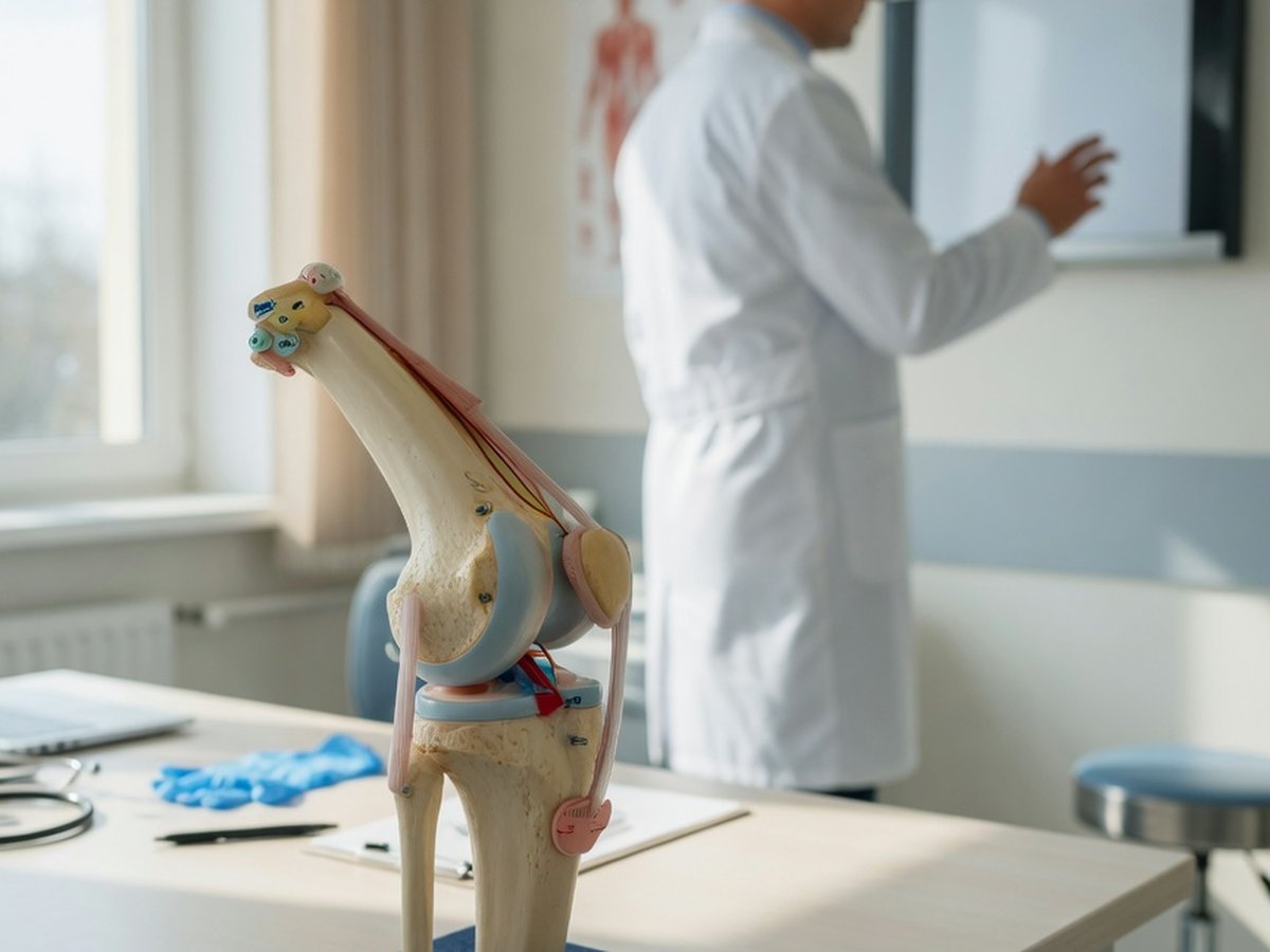
3D illustration of the human knee joint with AI analysis highlights — ReadYourLab AI knee MRI analysis

Why knee MRI reports are so hard to read

A knee MRI report is written for the orthopedic surgeon who referred you, not for you. It's full of terms like "grade 2 signal within the posterior horn of the medial meniscus" and "chondral fibrillation" — with little indication of what actually needs attention.

As with the spine, a lot of what shows up on a knee MRI is ordinary wear. Meniscus signal changes and cartilage thinning are frequently seen in people with no knee pain, and become more common with age. A finding on the scan is not automatically the source of your symptoms.

ReadYourLab reads your images with Google's medical AI and rewrites the report in plain language: what each finding is, how common it is, and which ones typically warrant a conversation with your doctor. It's an informed first look to help you prepare — not a diagnosis.

Common knee MRI findings — explained

The terms that show up most often on knee reports, and what they usually mean.

Meniscus tear

Depends on type & age

The meniscus is the C-shaped shock absorber in the knee. Tears range from small degenerative fraying — very common with age and often symptom-free — to acute tears from a twist. Whether one needs treatment depends on the type, your symptoms, and your activity, not the word "tear" alone.

ACL / PCL injury

Ask your doctor

The cruciate ligaments stabilize the knee. A full ACL tear usually follows a specific injury and often needs orthopedic evaluation, especially if the knee feels unstable. Partial or old, healed injuries are read differently.

Chondromalacia / cartilage wear

Very common

Softening or thinning of the smooth cartilage on the joint surfaces, graded 1–4. Low grades are extremely common and often painless; it's an early marker of wear rather than a diagnosis by itself.

Full explanation

Osteoarthritis changes

Common with age

Bony spurs (osteophytes), joint-space narrowing, and cartilage loss. Common with age and often gradual; severity on imaging doesn't always match how much pain you feel.

Full explanation

Joint effusion

Context matters

Extra fluid in the knee joint. A small amount can be normal; a larger effusion suggests irritation, inflammation, or a response to another finding, and is read together with the rest of the scan.

Baker's (popliteal) cyst

Usually benign

A fluid-filled pocket behind the knee that often connects with joint swelling. Common, usually harmless, and frequently linked to other changes inside the joint rather than a problem on its own.

Full explanation

Bone marrow edema

Read in context

A bright signal in the bone that can reflect a bruise, stress reaction, or arthritis-related change. Its meaning depends heavily on the surrounding findings and your history.

Bursitis / tendinosis

Often manageable

Irritation of a fluid-cushioning bursa or wear in a tendon. Common causes of localized knee pain that are frequently managed without surgery.

Descriptions here are general education, not a reading of your specific scan. What matters is how a finding lines up with your symptoms and exam — which is exactly what your doctor weighs.

The grading scales in your knee report

Radiologists compress detail into short grades. These are the ones you're most likely to meet on a knee MRI — and what the numbers describe.

Chondromalacia grade (1–4)

Learn more

Describes how worn the joint cartilage looks, from mild softening (grade 1) to full-thickness loss exposing bone (grade 4). Low grades are common and often painless; the grade describes the cartilage, not your pain.

Kellgren–Lawrence grade (0–4)

Learn more

An overall osteoarthritis severity score based on joint-space narrowing and bony spurs. A higher grade means more visible wear — but imaging severity and symptoms don't always match.

Sample AI report excerpt

What your AI knee report looks like

A short excerpt from a real, anonymized knee analysis — rendered the way you'll see it.

Patient explanation

Knee MRI — Plain-Language Summary

Your scan shows joint swelling and wear-related changes, without a fresh fracture.

There is a moderate amount of fluid in the knee joint and degenerative changes with bony spurs. A likely Baker's cyst sits behind the knee, which often goes along with joint swelling.

Key points

  • Moderate joint fluid, which can occur with arthritis or inflammation.
  • Likely Baker's cyst behind the knee — a fluid pocket that often connects with joint swelling.
  • No definite acute fracture visible on this series.

Excerpt shown for illustration. Your report is generated from your own images.

How to analyze your knee MRI

From files on a disc to a plain-language report in a few minutes.

  1. 1 Select the folder with your knee MRI DICOM files (.dcm) — from a CD, USB stick, or your patient portal download.
  2. 2 Preview the slices in the free online viewer to confirm you've picked the right series.
  3. 3 Upload the series for AI analysis (a quick sign-up is needed for your first report).
  4. 4 Read your plain-language knee report in minutes, then ask follow-up questions about any finding.

Why ReadYourLab for your knee MRI

Built for patients, reviewed by a physician, honest about its limits.

Reads the whole 3D volume

The AI analyzes your full stack of slices as a 3D volume, understanding how findings connect across the joint — not one flat image at a time.

Physician-reviewed & transparent

Our approach is medically reviewed, we tell you which model read your scan, and we're clear that this is an educational first look, not a diagnosis.

Private by design

Your images are encrypted in transit and at rest, never sold, and you can delete them at any time. GDPR compliant.

Knee MRI analysis — frequently asked questions

Common questions about AI knee MRI analysis, meniscus and cartilage findings, and what your report means.

Can AI read my knee MRI?

Yes. Upload your knee MRI DICOM files and the AI produces a plain-language explanation of the findings — meniscus and ligament tears, cartilage wear, joint fluid, and Baker's cysts — in a few minutes. It's an educational first look to help you understand your report and prepare questions, not a diagnosis or a replacement for your radiologist or orthopedic surgeon.

What does chondromalacia or a cartilage grade mean?

Chondromalacia is softening or thinning of the joint cartilage, graded 1 to 4. Low grades are very common and often painless — an early marker of wear rather than a diagnosis on their own. Our report explains your grade in plain language and links to a fuller explanation.

Is my knee MRI kept private?

Yes. Your images are encrypted in transit and at rest, are never shared with third parties or sold, and you can delete them at any time. ReadYourLab is GDPR compliant.

Is this a diagnosis or a replacement for my doctor?

No. ReadYourLab is an educational tool, not a medical device, and does not provide a diagnosis. The AI can be wrong — it may miss real findings or describe ones that aren't clinically important. Always review your results with your own doctor, and seek prompt care for a knee that is locked, gives way, or is severely swollen and painful after an injury.

Understand your knee MRI today

Your first AI report is free — no credit card required.

Analyze My Knee MRI