Foot & Ankle X-ray

Understand your foot or ankle X-ray — in language that actually makes sense

Upload your foot or ankle X-ray — a file or even a phone photo of a printed film — and get a clear, plain-language explanation of what the findings mean: fractures, heel spurs, bunions, arthritis, and more.

GDPR compliant Encrypted Delete anytime
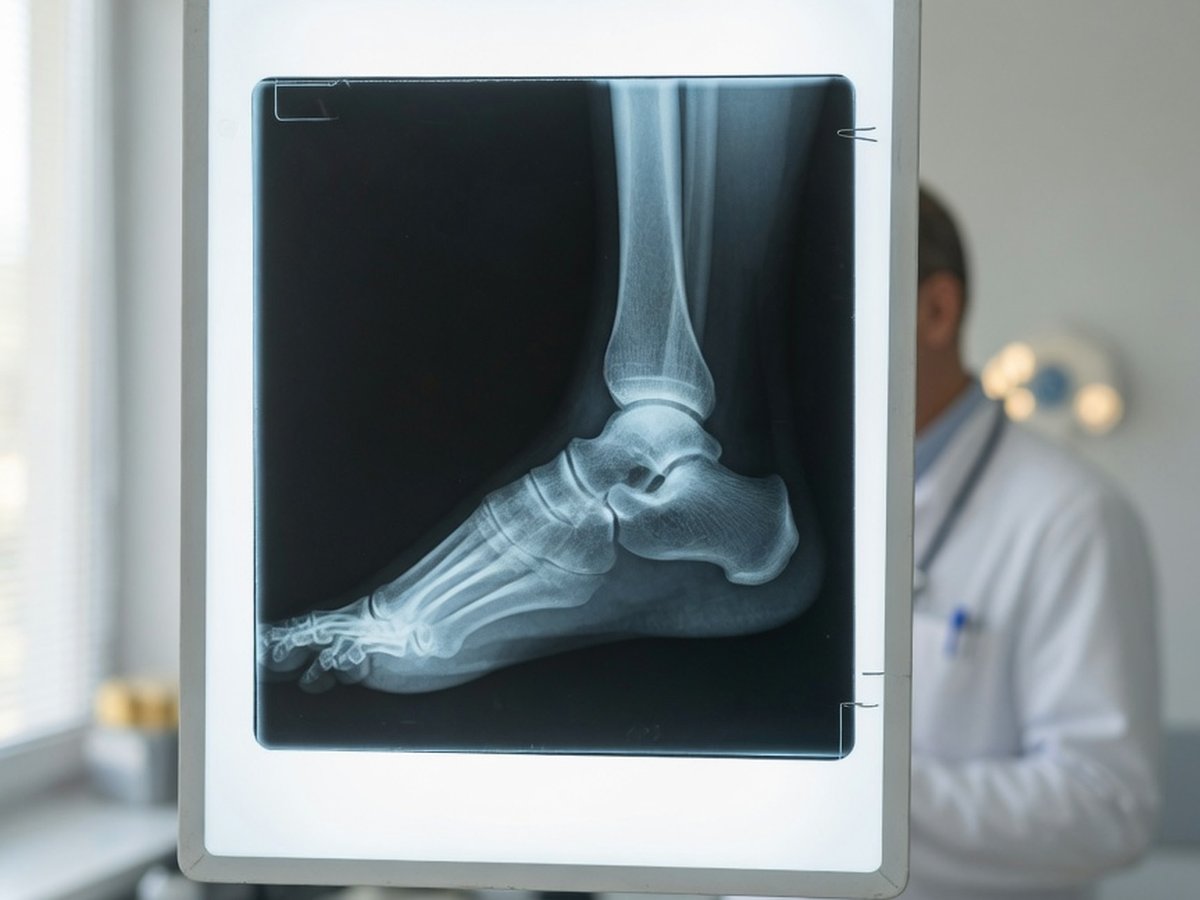
Foot and ankle X-ray film on a lightbox in a clinic — ReadYourLab AI foot and ankle X-ray interpretation

Why foot and ankle X-ray reports are hard to read

The foot and ankle contain 26 bones and dozens of joints, so the report reads like a roll call of unfamiliar names — malleolus, calcaneus, talus, the metatarsals — with terse notes about each.

A lot of what gets reported is common and often painless: a heel spur is frequently an incidental finding, and mild arthritis is nearly universal with age. Meanwhile some real injuries — hairline and stress fractures especially — can be invisible on an early film.

ReadYourLab reads your image with medical AI and rewrites the report in plain language: what each finding is, how common it is, and which ones typically warrant a conversation with your doctor. It's an informed first look — not a diagnosis.

Common foot & ankle X-ray findings — explained

The terms that show up most often on foot and ankle X-ray reports, and what they usually mean.

Ankle (malleolar) fracture

Needs attention

A break of the bony bumps on either side of the ankle, usually after a twist or fall. Whether it needs a cast or surgery depends on which side is broken and whether the ankle is stable.

Full explanation

Avulsion fracture

Often minor

A small chip of bone pulled off by a ligament or tendon during a sprain. Common in ankle injuries and frequently treated much like a bad sprain rather than a major break.

Full explanation

Stress fracture

Can be invisible early

A hairline break from repetitive load rather than one injury. Often not visible on an early X-ray and only appears weeks later as it heals — so a normal film doesn't rule it out.

Full explanation

Heel spur (calcaneal)

Usually incidental

A bony point at the heel where tissue attaches. Very common and frequently found in people with no heel pain at all — the spur itself is rarely the cause of symptoms.

Full explanation

Plantar fasciitis changes

Clinically diagnosed

Signs of irritation where the sole's thick band attaches to the heel. This is diagnosed mostly from your symptoms and exam; the X-ray is often obtained to rule other things out.

Full explanation

Hallux valgus (bunion)

Common

The big toe angles toward the others and the joint at its base juts outward. Common, often gradual, and treated based on pain and footwear rather than on the X-ray angle alone.

Osteoarthritis changes

Common with age

Joint space narrowing and bony spurs, most often at the big-toe joint or the ankle. Common with age and frequently manageable without surgery.

Full explanation

Descriptions here are general education, not a reading of your specific X-ray. Always review your report with your doctor, and seek prompt care after an injury if you cannot bear weight, or if the foot or ankle is deformed, numb, or severely swollen.

Sample AI report excerpt

What your AI foot X-ray report looks like

A short excerpt from a real, anonymized ankle X-ray analysis — rendered the way you'll see it.

Patient explanation

Ankle X-ray — Plain-Language Summary

Your X-ray shows a small chip of bone off the outer ankle, with the joint itself well aligned.

There is a small avulsion fragment at the outer ankle bone, the kind commonly seen after a sprain where a ligament tugs a fleck of bone away. The ankle joint lines up normally and the larger bones look intact.

Key points

  • Small avulsion fragment at the outer ankle — a common sprain-related finding worth discussing with your doctor.
  • The ankle joint appears normally aligned, with no widening.
  • No other fracture is visible on this image — though early stress fractures can be hidden.

Excerpt shown for illustration. Your report is generated from your own image.

How to analyze your foot or ankle X-ray

From an image on your phone to a plain-language report in minutes.

  1. 1 Upload your foot or ankle X-ray — a JPEG, PNG, or DICOM file, or simply a phone photo of a printed film.
  2. 2 Optionally add your age, sex, and any symptoms — clinical context measurably sharpens the interpretation.
  3. 3 Run the analysis (a quick sign-up is needed for your first report).
  4. 4 Read your plain-language report in minutes, then ask follow-up questions about any finding.

Why ReadYourLab for your foot X-ray

Built for patients, honest about its limits.

Works with a phone photo

No special software or disc needed. Upload a JPEG, PNG, or DICOM — a clear photo of a printed film works too.

Severity-coded, plain language

Findings are explained in words you can actually use, flagged by how much attention they typically warrant, with a chat for follow-up questions.

Private by design

Your image is encrypted in transit and at rest, never sold, and you can delete it at any time. GDPR compliant.

Foot & ankle X-ray analysis — frequently asked questions

Common questions about AI foot and ankle X-ray analysis, uploads, and privacy.

Can AI read my foot or ankle X-ray?

Yes. Upload your foot or ankle X-ray and the AI produces a plain-language explanation of the findings — fractures, heel spurs, bunions, arthritis, and alignment — in a few minutes, with severity-coded findings and a summary. It's an educational first look to help you understand your report and prepare questions, not a diagnosis or a replacement for your radiologist.

What file formats can I upload — can I photograph a printed film?

You can upload a JPEG, PNG, or a single-image DICOM (.dcm) file. A clear phone photo of a printed X-ray film works too — just capture the whole film, in focus, with as little glare and reflection as possible.

Can the AI miss a fracture?

Yes. Stress fractures and some hairline breaks are genuinely invisible on an early X-ray and are missed by radiologists too, sometimes only showing up weeks later as they heal. A report that finds nothing is not proof that nothing is broken. If you can't bear weight or the pain persists after an injury, see a doctor regardless of what any analysis says.

Is my X-ray kept private?

Yes. Your image is encrypted in transit and at rest, is never shared with third parties or sold, and you can delete it at any time. ReadYourLab is GDPR compliant.

Is this a diagnosis or a replacement for my doctor?

No. ReadYourLab is an educational tool, not a medical device, and does not provide a diagnosis. The AI can be wrong — it may miss real findings or describe ones that aren't clinically important. Always review your results with your own doctor, and seek prompt care if you cannot bear weight or the foot or ankle is deformed or numb.

Understand your foot X-ray today

Your first AI report is free — no credit card required.

Analyze My Foot X-ray