Shoulder MRI

Understand your shoulder MRI — in language that actually makes sense

Upload your shoulder MRI and get a clear, plain-language explanation of what the findings mean — rotator cuff tendinosis and tears, labral and SLAP tears, bursitis, AC joint wear, and frozen shoulder — so you know what to ask before your appointment.

GDPR compliant Encrypted Delete anytime
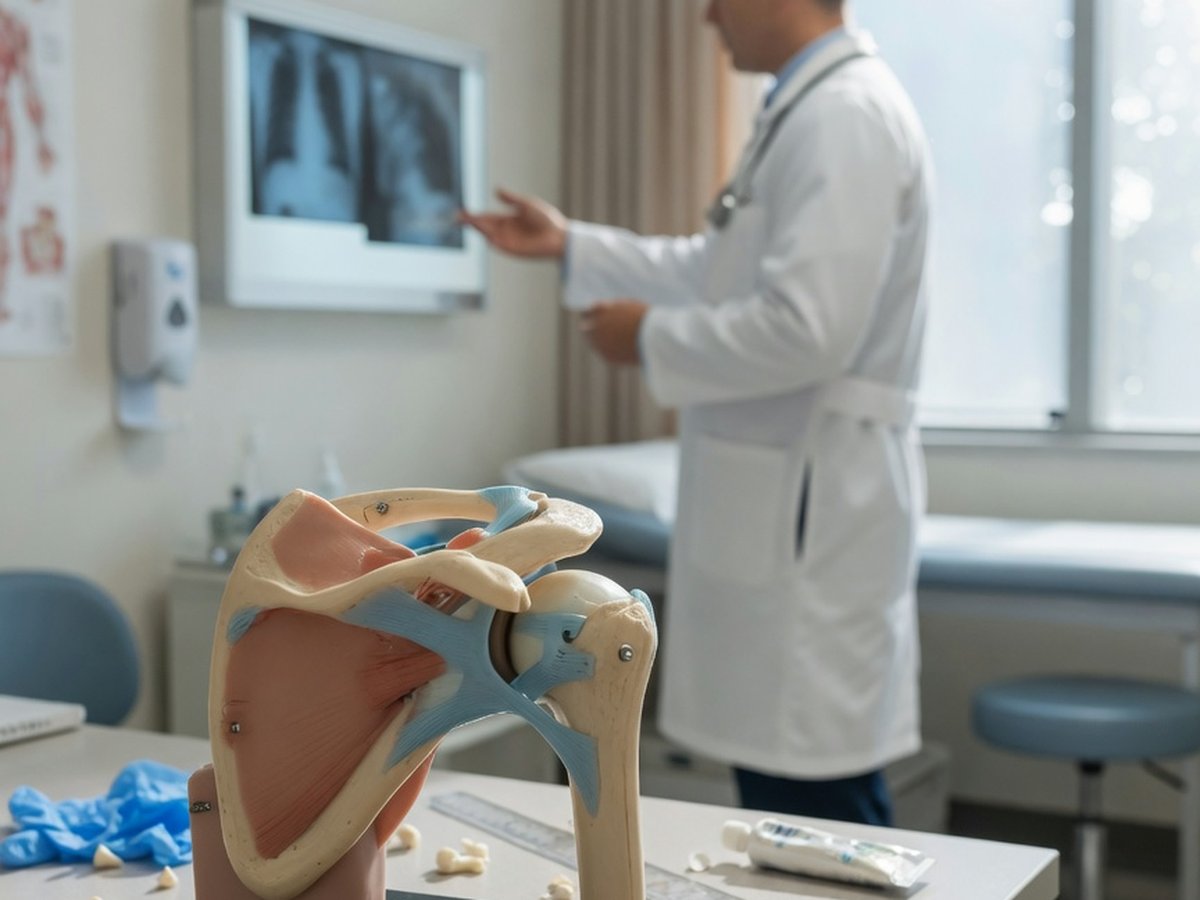
3D illustration of the human shoulder joint with AI analysis highlights — ReadYourLab AI shoulder MRI analysis

Why shoulder MRI reports are so hard to read

A shoulder MRI report is written for the orthopedic surgeon, packed with terms like "supraspinatus tendinosis with a low-grade articular-surface partial tear" and "superior labral fraying." It rarely tells you, in plain words, whether you're looking at normal wear or a real problem.

As with other joints, much of what shows up on a shoulder MRI is age-related change. Rotator cuff and labral wear are frequently seen in people with no shoulder pain, and become more common with each decade. A finding on the scan is not automatically the cause of your symptoms.

ReadYourLab reads your images with Google's medical AI and rewrites the report in plain language: what each finding is, how common it is, and which ones typically warrant a conversation with your doctor. It's an informed first look to help you prepare — not a diagnosis.

Common shoulder MRI findings — explained

The terms that show up most often on shoulder reports, and what they usually mean.

Rotator cuff tendinosis

Very common

Wear and thickening of a rotator cuff tendon (most often the supraspinatus) without a full tear. Extremely common with age and often painless; it sits at the mild end of the cuff-wear spectrum.

Full explanation

Rotator cuff tear (partial / full-thickness)

Depends on type

A tear in one of the cuff tendons, described as partial- or full-thickness. Small partial tears are common and often managed without surgery; full-thickness tears with weakness are more likely to prompt an orthopedic discussion. The word "tear" alone doesn't dictate treatment.

Full explanation

Subacromial–subdeltoid bursitis

Often manageable

Irritation and fluid in the bursa that cushions the cuff beneath the acromion. A common cause of shoulder pain that frequently responds to non-surgical treatment.

Full explanation

Labral tear (including SLAP)

Context matters

A tear of the cartilage rim that deepens the shoulder socket; a SLAP tear involves its upper part. Some are degenerative and symptom-free, others follow an injury and cause catching or instability — the pattern and your symptoms guide what to do.

Full explanation

AC joint arthrosis

Common with age

Wear of the acromioclavicular joint at the top of the shoulder, with bony spurs. Very common on imaging, often without symptoms, and only sometimes the source of pain.

Full explanation

Adhesive capsulitis (frozen shoulder)

Usually recovers

Thickening and inflammation of the joint capsule causing stiffness and pain. Often diagnosed clinically as much as on MRI, and typically improves over time with therapy.

Full explanation

Calcific tendinopathy

Often treatable

Calcium deposits within a rotator cuff tendon that can flare up painfully. Common, frequently self-limiting, and manageable with targeted treatments when symptomatic.

Full explanation

Impingement / acromion shape

Read in context

A hooked or low-lying acromion can crowd the cuff (impingement). It's a mechanical description that matters mainly when it lines up with pain when you raise the arm.

Full explanation

Descriptions here are general education, not a reading of your specific scan. What matters is how a finding lines up with your symptoms and exam — which is exactly what your doctor weighs.

How your shoulder report describes severity

The shoulder has no single number scale like the spine, but reports use a consistent vocabulary for how worn or torn a tendon is.

The cuff-wear spectrum: tendinosis → partial → full-thickness

Learn more

Rotator cuff findings sit on a spectrum from tendinosis (wear, no tear), to a partial-thickness tear (part of the tendon), to a full-thickness tear (all the way through). Where a finding sits on that line — plus any weakness — matters far more than the word "tear" alone.

Acromion type (I–III)

Learn more

Radiologists sometimes classify the acromion's shape from flat (Type I) to hooked (Type III). A more hooked shape can be associated with impingement, but on its own it's a description, not a diagnosis.

Sample AI report excerpt

What your AI shoulder report looks like

A short excerpt from a real, anonymized shoulder MRI analysis — rendered the way you'll see it.

Patient explanation

Shoulder MRI — Plain-Language Summary

Your scan shows rotator cuff wear with a small partial tear, plus bursal irritation — commonly managed without surgery first.

The supraspinatus tendon shows tendinosis with a low-grade partial tear on its under-surface, and there is fluid in the subacromial bursa. The rotator cuff is otherwise intact, with no full-thickness tear or muscle wasting.

Key points

  • Supraspinatus tendinosis with a small partial-thickness tear — often managed without surgery at first.
  • Subacromial bursitis, which can contribute to pain when raising the arm.
  • No full-thickness tear and no muscle wasting on this series.

Excerpt shown for illustration. Your report is generated from your own images.

How to analyze your shoulder MRI

From files on a disc to a plain-language report in a few minutes.

  1. 1 Select the folder with your shoulder MRI DICOM files (.dcm) — from a CD, USB stick, or your patient portal download.
  2. 2 Preview the slices in the free online viewer to confirm you've picked the right series.
  3. 3 Upload the series for AI analysis (a quick sign-up is needed for your first report).
  4. 4 Read your plain-language shoulder report in minutes, then ask follow-up questions about any finding.

Why ReadYourLab for your shoulder MRI

Built for patients, reviewed by a physician, honest about its limits.

Reads the whole 3D volume

The AI analyzes your full stack of slices as a 3D volume, understanding how findings connect across the joint — not one flat image at a time.

Physician-reviewed & transparent

Our approach is medically reviewed, we tell you which model read your scan, and we're clear that this is an educational first look, not a diagnosis.

Private by design

Your images are encrypted in transit and at rest, never sold, and you can delete them at any time. GDPR compliant.

Shoulder MRI analysis — frequently asked questions

Common questions about AI shoulder MRI analysis, rotator cuff findings, and what your report means.

Can AI read my shoulder MRI?

Yes. Upload your shoulder MRI DICOM files and the AI produces a plain-language explanation of the findings — rotator cuff tendinosis and tears, labral and SLAP tears, bursitis, and AC joint wear — in a few minutes. It's an educational first look to help you understand your report and prepare questions, not a diagnosis or a replacement for your radiologist or orthopedic surgeon.

Is my shoulder MRI kept private?

Yes. Your images are encrypted in transit and at rest, are never shared with third parties or sold, and you can delete them at any time. ReadYourLab is GDPR compliant.

Is this a diagnosis or a replacement for my doctor?

No. ReadYourLab is an educational tool, not a medical device, and does not provide a diagnosis. The AI can be wrong — it may miss real findings or describe ones that aren't clinically important. Always review your results with your own doctor, especially after an injury or if the shoulder is weak, unstable, or severely limited.

Understand your shoulder MRI today

Your first AI report is free — no credit card required.

Analyze My Shoulder MRI