Chest X-ray

Understand your chest X-ray — in language that actually makes sense

Upload your chest X-ray — a file or even a phone photo of a printed film — and get a clear, plain-language explanation of what the findings mean: consolidation, pleural fluid, heart size, nodules, and the terms that sound worse than they are.

GDPR compliant Encrypted Delete anytime
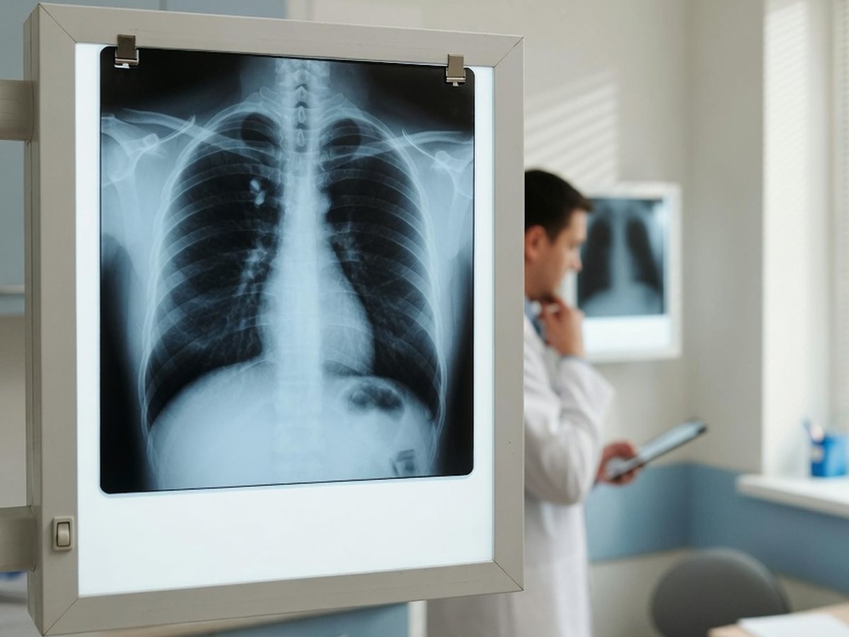
Chest X-ray film on a lightbox in a clinic — ReadYourLab AI chest X-ray interpretation

Why chest X-ray reports are so hard to read

The chest X-ray is the most common imaging test in medicine — and its report is written in a compressed shorthand meant for the referring doctor: "patchy opacity at the left base, blunting of the costophrenic angle, cardiomediastinal silhouette within normal limits."

Many of those phrases describe things that are normal, minor, or expected — a slightly prominent vessel, a bit of scarring, a heart that only looks large because of how the film was taken. Others genuinely need follow-up. The report rarely tells you which is which.

ReadYourLab reads your image with medical AI and rewrites it in plain language: what each finding is, how common it is, and which ones are worth a prompt conversation with your doctor. It's an informed first look to help you prepare — not a diagnosis.

Common chest X-ray findings — explained

The terms that show up most often on chest X-ray reports, and what they usually mean.

Consolidation / opacity

Often infection

An area of lung that looks white because air has been replaced by fluid, pus, or cells. It is the classic appearance of pneumonia, but can also follow other causes. It is usually treated and then re-imaged to confirm it clears.

Full explanation

Pleural effusion

Cause matters

Fluid collected between the lung and the chest wall. Small amounts are common after infection or surgery; larger ones prompt a search for the cause. It's the underlying reason, not the fluid itself, that guides treatment.

Full explanation

Costophrenic angle blunting

Often minor

The sharp corner where the diaphragm meets the ribs looks rounded off. It usually means a small amount of pleural fluid or old scarring — frequently a minor, incidental observation.

Full explanation

Atelectasis

Very common

A small area of lung that hasn't fully inflated, so it appears denser. Extremely common — especially after surgery, a deep breath not taken, or lying down — and usually resolves on its own.

Full explanation

Cardiomegaly (enlarged heart)

Read in context

The heart shadow looks wider than expected. Sometimes real, but often exaggerated by the technique — a front-to-back (AP) portable film magnifies the heart. It's typically confirmed with a proper film or an echocardiogram.

Full explanation

Pulmonary nodule

Usually followed up

A small round spot in the lung. Most are benign — old scars or healed infection — but because a minority are not, they're commonly followed with a CT or a repeat film to check for change over time.

Full explanation

Hyperinflation / emphysema

Chronic finding

The lungs look over-expanded with flattened diaphragms, a pattern seen in COPD and long-term smoking. It's a chronic, gradual change rather than an acute problem.

Full explanation

Aortic calcification / tortuosity

Common with age

The wall of the aorta shows calcium, or the vessel looks more curved than it once did. Both are common with age and blood-pressure history, and are usually noted rather than acted on directly.

Full explanation

Descriptions here are general education, not a reading of your specific X-ray. Always review your report with your doctor, and seek urgent care for severe breathlessness, chest pain, or coughing up blood.

The measurement in your chest report

Chest X-rays don't use a numbered severity scale the way joints do, but one ratio comes up often.

Cardiothoracic ratio (CTR)

Learn more

Compares the width of the heart to the width of the chest. Above roughly half is often flagged as an enlarged heart — but the number is only reliable on a properly taken film, and a portable or AP view routinely overstates it.

Sample AI report excerpt

What your AI chest X-ray report looks like

A short excerpt from a real, anonymized chest X-ray analysis — rendered the way you'll see it.

Patient explanation

Chest X-ray — Plain-Language Summary

Your X-ray shows a patch at the left lung base that fits with infection, and no fluid around the lungs.

There is a hazy area in the lower left lung of the kind commonly seen with pneumonia. The heart size looks normal for this view, and the edges where the lungs meet the diaphragm are sharp, which argues against a build-up of fluid.

Key points

  • Patchy opacity at the left lung base — a pattern that commonly reflects infection and is usually re-checked after treatment.
  • Heart size appears within normal limits on this view.
  • No pleural effusion and no collapsed lung seen on this image.

Excerpt shown for illustration. Your report is generated from your own image.

How to analyze your chest X-ray

From an image on your phone to a plain-language report in minutes.

  1. 1 Upload your chest X-ray — a JPEG, PNG, or DICOM file, or simply a phone photo of a printed film.
  2. 2 Optionally add your age, sex, and any symptoms — clinical context measurably sharpens the interpretation.
  3. 3 Run the analysis (a quick sign-up is needed for your first report).
  4. 4 Read your plain-language report in minutes, then ask follow-up questions about any finding.

Why ReadYourLab for your chest X-ray

Built for patients, honest about its limits.

Works with a phone photo

No special software or disc needed. Upload a JPEG, PNG, or DICOM — a clear photo of a printed film works too.

Severity-coded, plain language

Findings are explained in words you can actually use, flagged by how much attention they typically warrant, with a chat for follow-up questions.

Private by design

Your image is encrypted in transit and at rest, never sold, and you can delete it at any time. GDPR compliant.

Chest X-ray analysis — frequently asked questions

Common questions about AI chest X-ray analysis, uploads, and privacy.

Can AI read my chest X-ray?

Yes. Upload your chest X-ray and the AI produces a plain-language explanation of the findings — consolidation, pleural fluid, heart size, nodules, and more — in a few minutes, with severity-coded findings and a summary. It's an educational first look to help you understand your report and prepare questions, not a diagnosis or a replacement for your radiologist.

What file formats can I upload — can I photograph a printed film?

You can upload a JPEG, PNG, or a single-image DICOM (.dcm) file. A clear phone photo of a printed X-ray film works too — just capture the whole film, in focus, with as little glare and reflection as possible.

Is my X-ray kept private?

Yes. Your image is encrypted in transit and at rest, is never shared with third parties or sold, and you can delete it at any time. ReadYourLab is GDPR compliant.

Is this a diagnosis or a replacement for my doctor?

No. ReadYourLab is an educational tool, not a medical device, and does not provide a diagnosis. The AI can be wrong — it may miss real findings or describe ones that aren't clinically important. Always review your results with your own doctor, and seek urgent care for severe breathlessness, chest pain, or coughing up blood.

Understand your chest X-ray today

Your first AI report is free — no credit card required.

Analyze My Chest X-ray