

Findings marked on your images
Tap a finding and the viewer jumps to the exact slice, with the area outlined.
Get a plain-language report on your CT or MRI scan in minutes, powered by frontier medical AI
Choose the folder containing all your CT or MRI scan files
…or drag and drop the folder here
Reading DICOM files...
0 files processed
Select an image series to preview
Click any series on the left to open the interactive viewer.
Preparing images...
Loading...
1 / 1
What you get
Clear findings, marked on your own images, and ready to share with your doctor.
Tap a finding and the viewer jumps to the exact slice, with the area outlined.

On CT scans, the major organs are detected and colored automatically, so you always know what you are looking at.

Turn your CT or MRI into a 3D model in the free viewer. Nothing to install, it runs in your browser.
Every finding is color-coded, so you see at a glance what needs attention and what is normal.
Each image series is read as a complete 3D stack, not one picture at a time.
Send your report and images as a read-only link or PDF document. No account is needed to open it.
Tap any underlined term, like LesionA general word for any spot or area of tissue that looks different from what surrounds it. On its own it does not mean cancer or anything specific.Read full definition →, in your report for a plain-language definition.
Ask anything about any report, in plain language, as often as you like.
Choose an option below to get started with your health analysis. Try any of them free!
Upload the scan files you received from your clinic and get a detailed, easy-to-understand report in minutes
Upload your X-ray image and get a clear, easy-to-understand explanation in minutes.
Upload your radiology report (PDF) and get a plain-language explanation. Also works for other lab notes.
Your first analysis is free — no credit card, no subscription.
Analysis packages from $30 · unused analyses never expire.
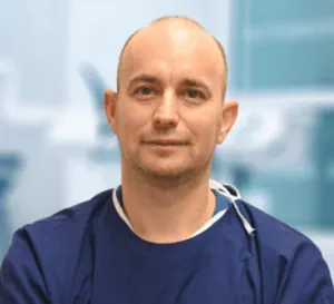
Dr. Zoltán Nyárády
Co-Founder · Oral & Maxillofacial Surgeon
MD, DMD, PhD
My specialty is oral surgery, dental implantation, and minimally invasive oral surgical procedures.
The greatest reward for me is when patients who once feared the dentist, oral surgeon, or implant procedures come back smiling for follow-up care — knowing I was able to ease their anxiety and show them that dental and oral surgery can be truly painless.
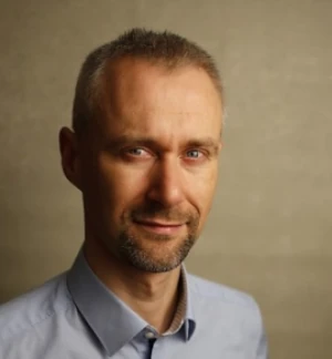
Peter Nemeth
Co-Founder
I spent most of my career building enterprise software products at ServiceNow, HP, and IBM. After a cancer diagnosis, I remember staring at a grey CT scan and feeling completely lost — with no idea what I was looking at. Using AI to finally understand what was happening in my own body was transformational.
I built ReadYourLab to give everyone access to the tool I wish I'd had during this time.
ReadYourLab makes state-of-the-art medical AI accessible to patients. We believe every patient deserves the ability to view and understand their own scans.
We believe the best way to make medical imaging more useful is to put it directly in the hands of patients. We want patients to be able to view, use, and understand their data in ways the traditional healthcare system simply doesn't allow. Patients should be able to decide how to engage with their own health and take control of their care in collaboration with their doctors.
AI-powered tools for explaining and visualizing the human body are a powerful way to give patients the freedom of information and medical education they deserve.
ReadYourLab runs on a state-of-the-art AI model, and state of the art still trails a board-certified radiologist. That is why every finding here is information to discuss with your doctor, not a diagnosis.
A better model is released almost every month, so your report is never tied to one vendor or one model name. Before any switch, the candidate reads real CT and MRI studies from our benchmark and is scored against the signed radiologist reports. Only a model that matches or beats the current one goes live.
We follow Radiology's Last Exam (RadLE), an independent benchmark of expert-level cases built by academic radiologists. Board-certified radiologists still score highest, ahead of every AI model, and general frontier models outscore smaller specialized medical models. We re-test whenever the leaderboard moves. Paper on arXiv · Live leaderboard
ReadYourLab gives you an AI-generated, plain-language walk-through of your images so you understand your scan before or after your doctor's appointment. It is not a substitute for a licensed radiologist or physician. Always consult your healthcare provider for medical decisions. AI vs. Radiologist — honest comparison.
Your scans are encrypted in transit and at rest. We never share your medical data with third parties. You can delete all your data with a single click at any time. HIPAA- and GDPR-aligned practices. Read our data security policy.
Common questions about AI-powered CT and MRI analysis, DICOM uploads, and our radiology reports.
Your first analysis is free. Your selected image series are kept while you sign up.
By continuing, you agree to the Terms and Conditions.
Already have an account? Need an account?
We sent a 6-digit code to . Enter it below to activate your account. Your selected image series are kept.
Didn't receive it? Check your spam folder or .
Your account is ready — you can manage your data anytime in your profile.
Date of birth and sex help the AI interpret your scans more accurately.
Your radiology report lets the Full Scan Report compare the AI findings with what your radiologist wrote. You can also add an earlier scan's report, lab results or other clinical notes — up to 3 PDFs. They are used for the Full Scan Report only.
Maximum of 3 documents attached.
Before you continue, please confirm each of the following:
Before you upload
Large scans can take a few minutes. Please keep this tab open until it's done.
No image series uploaded yet
1 / 1
Highlights organs detected by AI
Preparing 3D view
Drag to rotate · Scroll or pinch to cut through
Visualization is illustrative and not for clinical purposes.