Spine X-ray

Understand your spine X-ray — in language that actually makes sense

Upload your lumbar, cervical, or thoracic spine X-ray — a file or even a phone photo of a printed film — and get a clear, plain-language explanation: disc space narrowing, bone spurs, slippage, curvature, and fractures.

GDPR compliant Encrypted Delete anytime
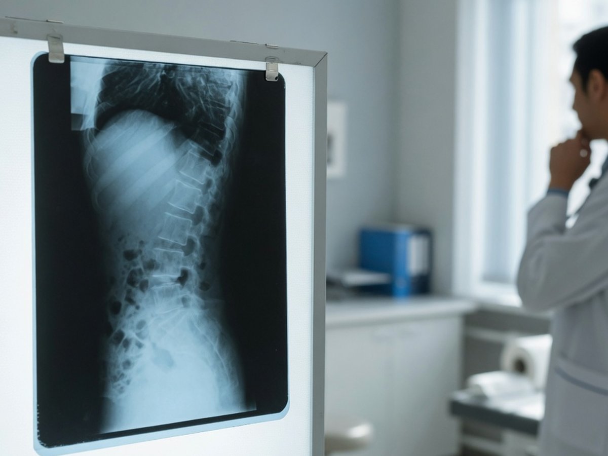
Spine X-ray film on a lightbox in a clinic — ReadYourLab AI spine X-ray interpretation

What a spine X-ray can and can't show

A spine X-ray shows the bones and their alignment — how the vertebrae stack up, how much space sits between them, and whether anything has slipped or collapsed. It cannot see your discs, nerves, or spinal cord directly. Those need an MRI.

That means terms like "disc space narrowing" and "osteophyte formation" describe age-related wear inferred from the bone. Such changes are found in a large share of adults with no back pain at all, and become more common every decade.

ReadYourLab reads your image with medical AI and rewrites the report in plain language: what each finding is, how common it is, and which ones typically warrant a conversation with your doctor. It's an informed first look — not a diagnosis.

Common spine X-ray findings — explained

The terms that show up most often on spine X-ray reports, and what they usually mean.

Disc space narrowing

Very common

The gap between two vertebrae looks reduced, which indirectly suggests the disc has lost height. Very common with age; the X-ray infers the disc's state from the space it leaves behind.

Full explanation

Spondylosis / osteophytes

Very common

Bony spurs along the vertebral edges — the spine's normal response to years of load. Common, frequently symptom-free, and usually described rather than treated.

Full explanation

Spondylolisthesis

Graded, ask your doctor

One vertebra has slipped forward relative to the one below. Often mild and stable; the grade and your symptoms determine whether anything is done.

Full explanation

Scoliosis / curvature

Measured by angle

A sideways curve of the spine, measured as a Cobb angle. Small curves are common and often just monitored; larger ones may prompt referral.

Full explanation

Compression fracture

Needs attention

A vertebra has lost height, typically from injury or weakened bone. Some are old and healed, some are new — telling them apart matters, and often needs your history or further imaging.

Full explanation

Facet joint arthrosis

Common with age

Wear of the small paired joints at the back of the spine. A frequent age-related finding that is read alongside your symptoms.

Full explanation

Osteopenia / reduced bone density

Discuss with your doctor

The bones look less dense than expected. A plain X-ray is an unreliable way to judge this — a dedicated DEXA bone-density scan is the proper test if it's raised.

Full explanation

Descriptions here are general education, not a reading of your specific X-ray. Always review your report with your doctor, and seek prompt care for leg weakness, loss of bladder or bowel control, numbness in the saddle area, or severe pain after an injury.

The measurements in your spine report

Two numbers come up often on spine X-rays — one for slippage, one for curvature.

Meyerding grade (I–IV)

Learn more

Rates how far one vertebra has slipped over the one below, in quarters. Grade I (up to a quarter) is the most common by far and is frequently managed without surgery.

Cobb angle

Learn more

Measures a sideways spinal curve in degrees. Roughly under 10° isn't considered scoliosis at all; larger angles guide whether monitoring or referral is appropriate.

Sample AI report excerpt

What your AI spine X-ray report looks like

A short excerpt from a real, anonymized lumbar spine X-ray analysis — rendered the way you'll see it.

Patient explanation

Lumbar Spine X-ray — Plain-Language Summary

Your X-ray shows common age-related wear in the lower back, with the vertebrae well aligned.

The space between the two lowest vertebrae looks reduced, with small bony spurs at the edges — a typical wear pattern. The vertebrae line up normally and none has lost height.

Key points

  • Disc space narrowing with marginal spurs at the lowest level — common, age-related wear.
  • Vertebrae are normally aligned, with no slippage seen.
  • No compression fracture and no loss of vertebral height on this image.

Excerpt shown for illustration. Your report is generated from your own image.

How to analyze your spine X-ray

From an image on your phone to a plain-language report in minutes.

  1. 1 Upload your spine X-ray — a JPEG, PNG, or DICOM file, or simply a phone photo of a printed film.
  2. 2 Optionally add your age, sex, and any symptoms — clinical context measurably sharpens the interpretation.
  3. 3 Run the analysis (a quick sign-up is needed for your first report).
  4. 4 Read your plain-language report in minutes, then ask follow-up questions about any finding.

Why ReadYourLab for your spine X-ray

Built for patients, honest about its limits.

Works with a phone photo

No special software or disc needed. Upload a JPEG, PNG, or DICOM — a clear photo of a printed film works too.

Severity-coded, plain language

Findings are explained in words you can actually use, flagged by how much attention they typically warrant, with a chat for follow-up questions.

Private by design

Your image is encrypted in transit and at rest, never sold, and you can delete it at any time. GDPR compliant.

Spine X-ray analysis — frequently asked questions

Common questions about AI spine X-ray analysis, uploads, and privacy.

Can AI read my spine X-ray?

Yes. Upload your lumbar, cervical, or thoracic spine X-ray and the AI produces a plain-language explanation of the findings — disc space narrowing, bone spurs, alignment and slippage, curvature, and fractures — in a few minutes, with severity-coded findings and a summary. It's an educational first look to help you understand your report and prepare questions, not a diagnosis or a replacement for your radiologist.

What file formats can I upload — can I photograph a printed film?

You can upload a JPEG, PNG, or a single-image DICOM (.dcm) file. A clear phone photo of a printed X-ray film works too — just capture the whole film, in focus, with as little glare and reflection as possible.

Can an X-ray show my discs or a pinched nerve?

Not directly — and neither can our analysis of one. X-rays show bone, so a disc's condition is only inferred from the space it leaves between vertebrae; nerves and the spinal cord are invisible on a plain film. Those are assessed with an MRI. If you have a spine MRI as well, you can upload it separately for a dedicated spine MRI analysis.

Is my X-ray kept private?

Yes. Your image is encrypted in transit and at rest, is never shared with third parties or sold, and you can delete it at any time. ReadYourLab is GDPR compliant.

Is this a diagnosis or a replacement for my doctor?

No. ReadYourLab is an educational tool, not a medical device, and does not provide a diagnosis. The AI can be wrong — it may miss real findings or describe ones that aren't clinically important. Always review your results with your own doctor, and seek prompt care for leg weakness, loss of bladder or bowel control, or numbness in the saddle area.

Understand your spine X-ray today

Your first AI report is free — no credit card required.

Analyze My Spine X-ray