Hand & Wrist X-ray

Understand your hand or wrist X-ray — in language that actually makes sense

Upload your hand or wrist X-ray — a file or even a phone photo of a printed film — and get a clear, plain-language explanation of what the findings mean: fractures, arthritis, joint changes, and the terms that sound worse than they are.

GDPR compliant Encrypted Delete anytime
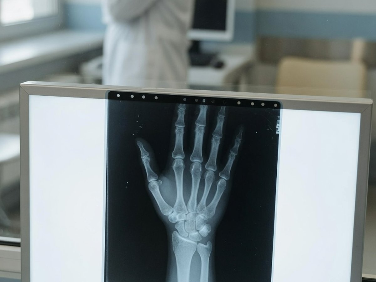
Hand and wrist X-ray film on a lightbox in a clinic — ReadYourLab AI hand and wrist X-ray interpretation

Why hand and wrist X-ray reports are hard to read

The hand and wrist pack dozens of small bones into a tiny space, so the report names structures most people have never heard of — scaphoid, lunate, the distal radius, the MCP, PIP and DIP joints — and describes them in a dense shorthand.

Much of what's reported is ordinary wear: arthritis at the base of the thumb or the finger-tip joints is very common with age and often manageable. But the wrist is also where a genuinely important fracture can be subtle — a scaphoid break can be nearly invisible on a first film.

ReadYourLab reads your image with medical AI and rewrites the report in plain language: what each finding is, how common it is, and which ones typically warrant a conversation with your doctor. It's an informed first look — not a diagnosis.

Common hand & wrist X-ray findings — explained

The terms that show up most often on hand and wrist X-ray reports, and what they usually mean.

Distal radius fracture (Colles)

Needs attention

A break of the forearm bone just above the wrist — the classic fall-on-an-outstretched-hand injury and the most common wrist fracture. Whether it needs setting depends on how the pieces sit.

Full explanation

Scaphoid fracture

Can be subtle

A break in a small wrist bone near the base of the thumb. Notorious for being invisible on the first X-ray. If your wrist hurts there after a fall, it's often treated as a fracture and re-imaged even when the film looks clear.

Full explanation

Osteoarthritis of the hand

Very common

Wear at the finger joints or the base of the thumb, showing as narrowing and spurs. Extremely common with age, often gradual, and usually managed without surgery.

Full explanation

Joint space narrowing

Very common

The gap between two bones looks thinner, indirectly suggesting worn cartilage. The main way arthritis shows up on a hand X-ray.

Full explanation

Osteophytes (bone spurs)

Very common

Small bony outgrowths at joint edges — a normal response to years of use. Often visible as the bumps people notice on their finger joints.

Full explanation

Erosions

Discuss with your doctor

Small areas where bone at a joint margin looks eaten away, a pattern that can point toward inflammatory arthritis rather than simple wear. This is one worth reviewing promptly with your doctor.

Reduced bone density

Unreliable on X-ray

The bones look less dense than expected. A plain X-ray is a poor way to judge this — a dedicated DEXA bone-density scan is the proper test if it's raised.

Full explanation

Soft tissue swelling

Descriptive

The soft tissues around a joint look puffier than normal. A non-specific sign that something is irritated, read together with the bones and your symptoms.

Descriptions here are general education, not a reading of your specific X-ray. Always review your report with your doctor, and seek prompt care after an injury — especially for wrist pain at the base of the thumb, an obvious deformity, or numbness.

Sample AI report excerpt

What your AI hand X-ray report looks like

A short excerpt from a real, anonymized wrist X-ray analysis — rendered the way you'll see it.

Patient explanation

Wrist X-ray — Plain-Language Summary

Your X-ray shows a break of the forearm bone just above the wrist, in good position.

There is a fracture of the lower end of the radius, the larger forearm bone, close to the wrist. The bone fragments appear to line up reasonably well, and the small wrist bones look intact on this view.

Key points

  • Fracture of the distal radius — this needs prompt medical assessment.
  • The fragments appear reasonably aligned on this view.
  • No definite scaphoid fracture is visible on this image — though early scaphoid breaks can be hidden.

Excerpt shown for illustration. Your report is generated from your own image.

How to analyze your hand or wrist X-ray

From an image on your phone to a plain-language report in minutes.

  1. 1 Upload your hand or wrist X-ray — a JPEG, PNG, or DICOM file, or simply a phone photo of a printed film.
  2. 2 Optionally add your age, sex, and any symptoms — clinical context measurably sharpens the interpretation.
  3. 3 Run the analysis (a quick sign-up is needed for your first report).
  4. 4 Read your plain-language report in minutes, then ask follow-up questions about any finding.

Why ReadYourLab for your hand X-ray

Built for patients, honest about its limits.

Works with a phone photo

No special software or disc needed. Upload a JPEG, PNG, or DICOM — a clear photo of a printed film works too.

Severity-coded, plain language

Findings are explained in words you can actually use, flagged by how much attention they typically warrant, with a chat for follow-up questions.

Private by design

Your image is encrypted in transit and at rest, never sold, and you can delete it at any time. GDPR compliant.

Hand & wrist X-ray analysis — frequently asked questions

Common questions about AI hand and wrist X-ray analysis, uploads, and privacy.

Can AI read my hand or wrist X-ray?

Yes. Upload your hand or wrist X-ray and the AI produces a plain-language explanation of the findings — fractures, arthritis, joint space narrowing, spurs, and soft-tissue changes — in a few minutes, with severity-coded findings and a summary. It's an educational first look to help you understand your report and prepare questions, not a diagnosis or a replacement for your radiologist.

What file formats can I upload — can I photograph a printed film?

You can upload a JPEG, PNG, or a single-image DICOM (.dcm) file. A clear phone photo of a printed X-ray film works too — just capture the whole film, in focus, with as little glare and reflection as possible.

Can the AI miss a fracture?

Yes. Some fractures — the scaphoid in the wrist especially — are genuinely hard to see on a first X-ray and can be missed by radiologists too, only becoming visible days later or on other imaging. A report that finds nothing is not proof that nothing is broken. If you have pain after an injury, see a doctor regardless of what any analysis says.

Is my X-ray kept private?

Yes. Your image is encrypted in transit and at rest, is never shared with third parties or sold, and you can delete it at any time. ReadYourLab is GDPR compliant.

Is this a diagnosis or a replacement for my doctor?

No. ReadYourLab is an educational tool, not a medical device, and does not provide a diagnosis. The AI can be wrong — it may miss real findings or describe ones that aren't clinically important. Always review your results with your own doctor, and seek prompt care after an injury, especially with an obvious deformity or numbness.

Understand your hand X-ray today

Your first AI report is free — no credit card required.

Analyze My Hand X-ray