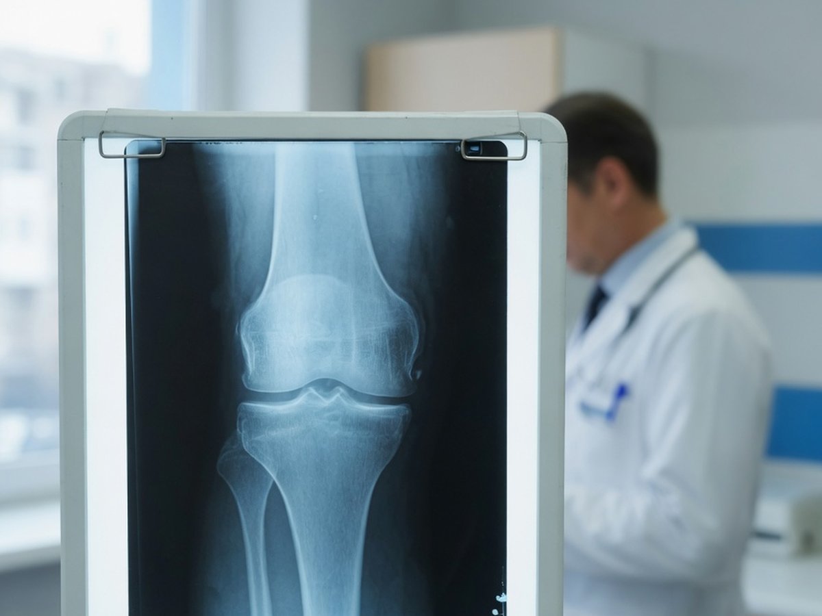
Knee X-ray

Understand your knee X-ray — in language that actually makes sense

Upload your knee X-ray — a file or even a phone photo of a printed film — and get a clear, plain-language explanation of what the findings mean: joint space narrowing, bone spurs, arthritis grading, and fractures.

GDPR compliant Encrypted Delete anytime
Knee X-ray film on a lightbox in a clinic — ReadYourLab AI knee X-ray interpretation

What a knee X-ray can and can't show

A knee X-ray shows bone beautifully and soft tissue barely at all. It's the right first test for arthritis, alignment, and fractures — but it cannot see your meniscus, your cruciate ligaments, or your cartilage directly. That's what an MRI is for.

So a report full of terms like "joint space narrowing" and "marginal osteophytes" is describing wear that is extremely common with age and often painless. How much a knee hurts and how bad it looks on film are famously poorly matched.

ReadYourLab reads your image with medical AI and rewrites the report in plain language: what each finding is, how common it is, and which ones typically warrant a conversation with your doctor. It's an informed first look — not a diagnosis.

Common knee X-ray findings — explained

The terms that show up most often on knee X-ray reports, and what they usually mean.

Joint space narrowing

Very common

The gap between the bones looks thinner, which indirectly suggests the cartilage cushion has worn down. It's the main way arthritis shows up on an X-ray, and it's very common with age.

Full explanation

Osteophytes (bone spurs)

Very common

Small bony outgrowths at the edges of the joint — the bone's normal response to years of load. Common, usually painless in themselves, and rarely removed just because they're visible.

Full explanation

Subchondral sclerosis

Wear-related

The bone just beneath the cartilage looks denser and whiter where it has taken extra load. A typical companion of joint wear rather than a separate problem.

Full explanation

Osteoarthritis changes

Common with age

The umbrella term when narrowing, spurs, and sclerosis appear together. Severity on film doesn't reliably predict pain — plenty of worn-looking knees feel fine, and vice versa.

Full explanation

Joint effusion

Context matters

Extra fluid in the knee, visible indirectly as swelling of the soft-tissue shadows. Suggests irritation or inflammation and is read alongside your symptoms.

Full explanation

Fracture

Needs attention

A break in the bone. Usually follows a specific injury and needs prompt medical care. Some hairline fractures are subtle on a first film and only become visible later or on other imaging.

Full explanation

Alignment (varus / valgus)

Descriptive

Whether the leg tilts bow-legged (varus) or knock-kneed (valgus). It describes how load is distributed through the knee and often accompanies one-sided wear.

Descriptions here are general education, not a reading of your specific X-ray. What matters is how a finding lines up with your symptoms and exam — which is exactly what your doctor weighs.

The grading scale in your knee report

Knee X-rays have one widely used severity score, and it's the number you're most likely to see.

Kellgren–Lawrence grade (0–4)

Learn more

Rates osteoarthritis from none (0) to severe (4), based on joint-space narrowing and bony spurs. A higher grade means more visible wear — but imaging severity and how much pain you feel often don't match.

Sample AI report excerpt

What your AI knee X-ray report looks like

A short excerpt from a real, anonymized knee X-ray analysis — rendered the way you'll see it.

Patient explanation

Knee X-ray — Plain-Language Summary

Your X-ray shows moderate wear on the inner side of the knee, with no fracture.

The gap between the bones is reduced on the inner (medial) side, with small bony spurs at the joint edges — a common pattern of age-related wear. The bones themselves look intact.

Key points

  • Moderate joint space narrowing on the inner side of the knee, with marginal bone spurs — a typical arthritis pattern.
  • No fracture and no dislocation seen on this image.
  • Bone density appears unremarkable for this view.

Excerpt shown for illustration. Your report is generated from your own image.

How to analyze your knee X-ray

From an image on your phone to a plain-language report in minutes.

  1. 1 Upload your knee X-ray — a JPEG, PNG, or DICOM file, or simply a phone photo of a printed film.
  2. 2 Optionally add your age, sex, and any symptoms — clinical context measurably sharpens the interpretation.
  3. 3 Run the analysis (a quick sign-up is needed for your first report).
  4. 4 Read your plain-language report in minutes, then ask follow-up questions about any finding.

Why ReadYourLab for your knee X-ray

Built for patients, honest about its limits.

Works with a phone photo

No special software or disc needed. Upload a JPEG, PNG, or DICOM — a clear photo of a printed film works too.

Severity-coded, plain language

Findings are explained in words you can actually use, flagged by how much attention they typically warrant, with a chat for follow-up questions.

Private by design

Your image is encrypted in transit and at rest, never sold, and you can delete it at any time. GDPR compliant.

Knee X-ray analysis — frequently asked questions

Common questions about AI knee X-ray analysis, uploads, and privacy.

Can AI read my knee X-ray?

Yes. Upload your knee X-ray and the AI produces a plain-language explanation of the findings — joint space narrowing, bone spurs, arthritis grading, alignment, and fractures — in a few minutes, with severity-coded findings and a summary. It's an educational first look to help you understand your report and prepare questions, not a diagnosis or a replacement for your radiologist.

What file formats can I upload — can I photograph a printed film?

You can upload a JPEG, PNG, or a single-image DICOM (.dcm) file. A clear phone photo of a printed X-ray film works too — just capture the whole film, in focus, with as little glare and reflection as possible.

Can an X-ray show my meniscus or ligaments?

No — and neither can our analysis of one. X-rays show bone; the meniscus, cruciate ligaments, and cartilage are soft tissue and are effectively invisible on a plain film. Those are assessed with an MRI. If you have an MRI as well, you can upload it separately for a dedicated knee MRI analysis.

Is my X-ray kept private?

Yes. Your image is encrypted in transit and at rest, is never shared with third parties or sold, and you can delete it at any time. ReadYourLab is GDPR compliant.

Is this a diagnosis or a replacement for my doctor?

No. ReadYourLab is an educational tool, not a medical device, and does not provide a diagnosis. The AI can be wrong — it may miss real findings or describe ones that aren't clinically important. Always review your results with your own doctor, especially after an injury or if the knee is locked, unstable, or severely swollen.

Understand your knee X-ray today

Your first AI report is free — no credit card required.

Analyze My Knee X-ray