Lumbar MRI: Disc Bulges, Stenosis, and Leg Symptoms
Plain-language guide to lumbar MRI terms, nerve narrowing, sciatica, numbness, weakness, foot drop, and urgent red flags.
Why lumbar MRI reports can feel confusing
A lumbar MRI can be full of words that sound alarming: disc bulge, foraminal stenosis, lateral recess narrowing, cauda equina crowding, pars defect, or grade 1 slippage. Many people read these terms after having sciatica, numbness, weakness, foot slap, heavy legs, or trouble walking and wonder: Does this explain what I am feeling?
The honest answer is: sometimes, but not from the MRI wording alone. Spine MRI findings matter most when they match three things together: your symptoms, your physical exam, and the exact nerve pathway involved. This article is general education and is not a diagnosis or personal medical advice.
Common lumbar MRI terms in plain language
Disc bulge or protrusion
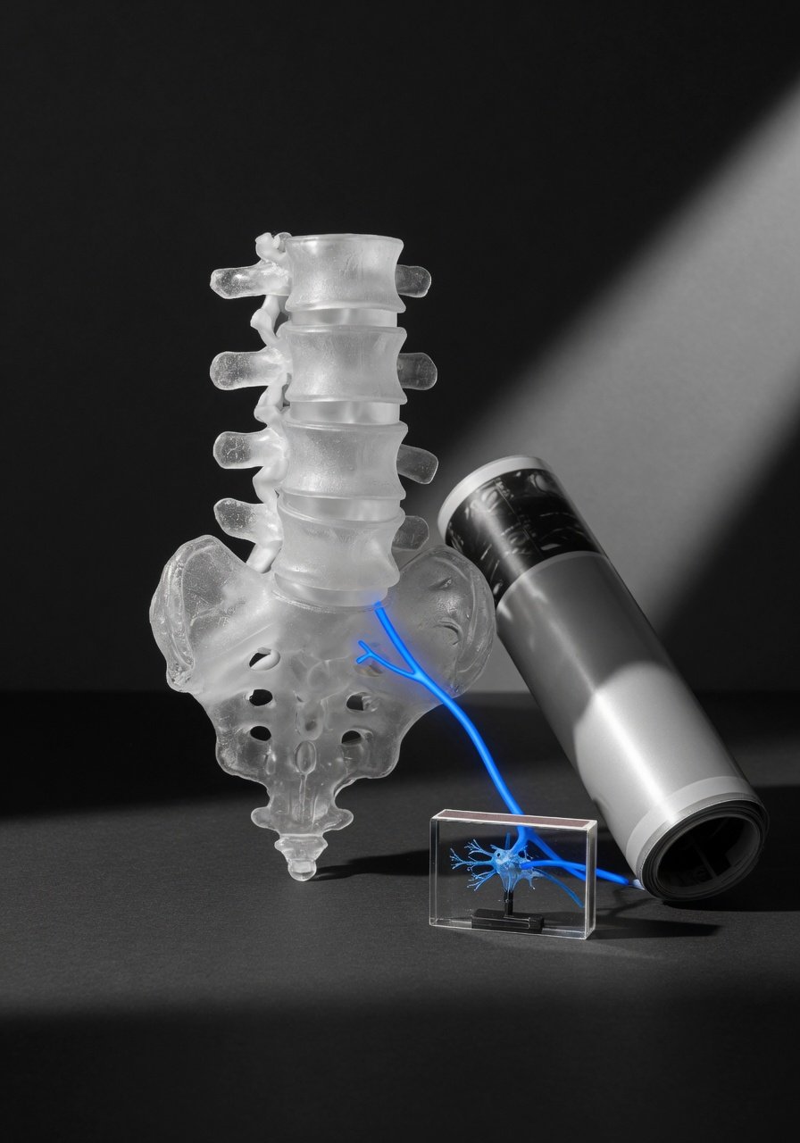
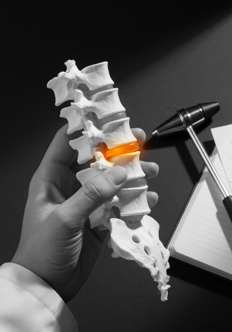
The discs are cushions between the bones of the spine. A disc bulge means the disc extends beyond its usual border. A protrusion is a more focal outpouching. These can touch or press on nearby nerve tissue, but many bulges are mild and may not cause symptoms.
Central canal stenosis
The central canal is the main tunnel that holds the nerve roots in the lower back. Stenosis means narrowing. Mild narrowing may be incidental. More severe narrowing can crowd the nerves and may cause leg heaviness, pain, numbness, or walking difficulty, especially when standing or walking.
Lateral recess narrowing
The lateral recess is a side hallway inside the spinal canal where a nerve root travels before exiting. Narrowing here can affect the traversing nerve root. For example, L4-L5 lateral recess narrowing often has the potential to affect the L5 nerve root.
Foraminal stenosis
The foramen is the side opening where a nerve exits the spine. Foraminal stenosis means that opening is narrowed. At L5-S1, foraminal stenosis can irritate or compress the exiting L5 nerve root. This can matter when symptoms follow an L5 pattern, such as pain or numbness down the outer leg toward the top of the foot, or weakness lifting the big toe or foot.
Pars defects and slippage
A pars defect, also called spondylolysis, is a stress-type defect in a small bridge of bone in the back of a vertebra. It can be old and stable, or it may contribute to back pain or spinal movement. Grade 1 anterolisthesis means one vertebra has slipped slightly forward. Flexion-extension X-rays are sometimes used because they are taken standing and bending, which may show movement that a lying-down MRI does not.
Cauda equina crowding
The cauda equina is the bundle of nerve roots at the bottom of the spinal canal. MRI reports may say the nerves are crowded when spinal fluid space around them is reduced. Crowding is not always an emergency, but severe compression with certain symptoms can be urgent.
Key idea: The scan describes anatomy. Your symptoms and neurologic exam help decide whether that anatomy is actually causing the problem.
How MRI levels relate to leg symptoms
Each lumbar nerve tends to serve a typical area of sensation and muscle strength. These patterns overlap, so they are not perfect, but they help clinicians match symptoms to imaging.
- L3 nerve: pain or numbness toward the front of the thigh and knee; possible trouble with hip flexion or knee extension.
- L4 nerve: symptoms toward the front/inner shin; possible weakness straightening the knee or lifting the foot inward.
- L5 nerve: pain, tingling, or numbness down the outer leg to the top of the foot or big toe; possible weakness lifting the foot or big toe, sometimes described as foot slap or scuffing.
- S1 nerve: symptoms down the back of the leg into the outer foot or sole; possible weakness pushing down, standing on tiptoe, or reduced ankle reflex.
This is why a report saying L4-L5 narrowing does not automatically mean the L4 nerve is affected. Depending on the location, L4-L5 narrowing may affect the L5 nerve in the lateral recess, while L4-L5 foraminal narrowing may affect the exiting L4 nerve.
Why right versus left matters
If pain, numbness, or weakness is mostly in the right leg, your clinician will look for a right-sided finding that matches the nerve pattern. A central disc bulge may narrow both sides or neither side meaningfully. A report may say bilateral narrowing, right foraminal stenosis, left lateral recess narrowing, or may not clearly state the side.
If the report does not specify laterality, it is reasonable for the treating clinician or radiologist to review the axial images, which are the cross-sectional slices. Those images often show whether the narrowing is right, left, central, or both.
Can mild MRI findings cause severe symptoms?
Sometimes symptoms feel much worse than the MRI sounds. There are several reasons this can happen. MRI is a still image taken while lying down. It may not show irritation that changes with standing, walking, extension, or bending. Inflammation around a nerve can also cause pain even without dramatic compression. On the other hand, some disc bulges and arthritis changes are common with aging and may not be the main cause of symptoms.
This is why exam findings are so important. A clinician may check reflexes, sensation, strength, walking pattern, ability to heel-walk or toe-walk, straight-leg raise, hip strength, and balance. They may also consider EMG and nerve conduction studies when weakness, foot drop, or numbness does not clearly match the MRI.
Sciatica, heavy legs, and neurogenic claudication
Sciatica usually refers to nerve-type pain traveling from the low back or buttock down the leg. It may feel sharp, burning, electric, or tingling. It often follows one nerve distribution.
Heavy legs or difficulty walking can occur with lumbar spinal stenosis, especially central canal stenosis. Some people feel worse when standing or walking and better when sitting or bending forward. This pattern is often called neurogenic claudication. However, leg heaviness can also come from hip problems, vascular circulation issues, medication effects, neurologic conditions, or general deconditioning, so it should not be assumed to be spine-related without evaluation.
Foot drop and weakness are different from pain alone
Pain is important, but weakness changes the urgency of the conversation. Foot drop, foot slap, frequent tripping, inability to lift the big toe, or trouble standing on tiptoe can suggest a motor nerve problem. These symptoms need prompt clinical attention because they may reflect nerve dysfunction, not just irritation.
Imaging can help look for a compressed nerve root, but weakness can also come from peripheral nerve injury, plexus injury, muscle injury, brain or spinal cord conditions, or functional neurologic patterns. The next step depends on the full medical story, exam, and timing.
Questions to ask about your lumbar MRI
- Which level is the main problem: L3-L4, L4-L5, or L5-S1?
- Is the narrowing central canal, lateral recess, foraminal, or more than one?
- Is it right-sided, left-sided, or bilateral?
- Which nerve root might be affected?
- Do my symptoms and exam match that nerve?
- Is there true weakness, reflex change, or sensory loss?
- Would standing X-rays, flexion-extension X-rays, EMG/NCS, or specialist review add useful information?
When to talk to your doctor
Talk with your doctor or a spine-trained clinician if back or leg symptoms persist, limit walking, include numbness or tingling, or do not match what you expected from the MRI report. Seek urgent medical care for new or worsening leg weakness, foot drop, numbness in the groin or saddle area, loss of bladder or bowel control, fever with severe back pain, or rapidly worsening symptoms. This article is general education only and cannot diagnose the cause of your symptoms.
Get AI-powered analysis of your CT or MRI scan
Upload your DICOM files and receive a clear, patient-friendly report in minutes.
Analyze my scan