
Synovitis on MRI: Does Joint-Lining Inflammation Mean Arthritis?
Mild synovitis on MRI does not automatically mean arthritis. Learn how joint fluid, tendon-sheath changes, injections, and surgery affect the findings.

Have your own scan or report? Get a clear, plain-language explanation in minutes.
Seeing “synovitis,” “synovial enhancement,” or “possible tenosynovitis” on an MRI report can raise an immediate question: Does this mean I have arthritis? Not necessarily. These terms describe changes in or around a joint, but they do not identify the cause on their own.
The distinction matters when a wrist MRI shows mild enhancement, or when a knee remains painful after surgery despite only a small amount of fluid. Symptoms, examination findings, the type of MRI, and recent procedures all help explain what the images mean.
This article provides general education, not a diagnosis or individual medical advice.
What is the joint lining?
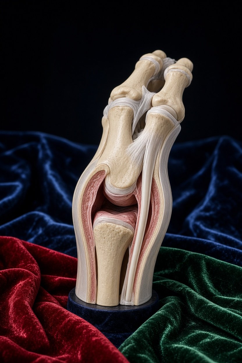
The synovium is a thin layer of tissue lining the inside of a joint capsule. It makes fluid that helps the joint move smoothly and supports the cartilage covering the ends of bones. The synovium is not the cartilage itself.
Synovitis means inflammation of this lining. On MRI, the lining may look thicker or become more visible after contrast is given into a vein. However, subtle changes can overlap with normal appearances or a mild, temporary response to irritation.
Synovitis, tenosynovitis, and joint fluid: three different findings
Synovitis: changes inside the joint capsule
Synovitis involves the joint lining. Radiologists consider its thickness, enhancement, distribution, and associated findings—not just whether fluid is present.
Tenosynovitis: changes around a tendon
Some tendons travel through sleeves called tendon sheaths, which also have a synovial lining. Inflammation of these sleeves is called tenosynovitis. In the wrist, “multicompartment tenosynovitis” means that several tendon-sheath compartments may be involved.
This is different from inflammation within the joint, although both can occur together. It is also different from a tendon tear: an irritated sheath does not mean the tendon is torn.
Joint fluid: not automatically inflammation
A small amount of lubricating fluid is normal. An increased amount is called an effusion. Fluid may accompany synovitis, but trace or small-volume fluid alone does not prove active inflammation or reveal its cause.
What does “mild synovial enhancement” mean?
After contrast is injected into a vein, tissues with a blood supply can become brighter on certain MRI images. This is called enhancement. Inflamed synovium may enhance, but normal synovium can enhance too.
For example, a wrist report may describe mild, smooth enhancement with minimal fluid and no bulky thickening of the lining. That appearance may reflect normal variation or low-grade synovitis. It is not the same as a diagnosis of rheumatoid arthritis.
Words such as “mild,” “possible,” and “nonspecific” communicate the degree of change or uncertainty. They do not mean that symptoms are imaginary, but they do limit what the scan can establish.
Does synovitis mean rheumatoid arthritis or infection?
No—synovitis is a finding, not a complete diagnosis. Possible causes include:
- Temporary irritation after injury or repetitive loading.
- Osteoarthritis, which can include episodes of joint-lining inflammation.
- Inflammatory arthritis, such as rheumatoid or psoriatic arthritis.
- Crystal-related inflammation, such as gout.
- Infection.
- Changes related to surgery or another procedure.
These causes can have overlapping MRI appearances. Mild enhancement or fluid alone does not establish any one of them.
Clinicians look at the broader pattern: which joints are affected, whether swelling is present, how long stiffness lasts, and whether there are other symptoms. Blood tests, ultrasound, or testing a sample of joint fluid may sometimes help. Suspected infection needs prompt clinical assessment; MRI alone cannot reliably settle that question.
Why a recent joint injection changes the interpretation
Not all contrast-enhanced scans are performed the same way. With an MR arthrogram, diluted contrast is injected directly into the joint before imaging. The injected fluid deliberately expands the joint, so the apparent fluid volume cannot simply be treated as a naturally occurring effusion.
Some contrast may also track along the needle path or leak into nearby soft tissues. This does not automatically prove a torn joint capsule. Around the wrist, procedure-related fluid may complicate the assessment of nearby tendon sheaths and possible tenosynovitis.
The radiologist needs to know what was injected, where, and when. A recent treatment injection, including a steroid injection, can also affect symptoms and inflammatory findings. The injection history belongs alongside the images.
After knee surgery: “Is there visible inflammation?”
That question is understandable when walking feels weak or squatting and going downstairs hurt. MRI can show signs that support inflammation, but it may not provide a definite yes-or-no answer.
After surgery, scar tissue and tendon thickening can persist without proving active inflammation. Small amounts of joint fluid and mild swelling in surrounding tissues are also nonspecific. Recent surgery can produce healing-related changes that overlap with inflammation; surgery years earlier provides a different context and should not automatically explain new symptoms.
Metal screws can distort MRI images, creating artifacts that obscure nearby cartilage, bone, and tendon attachments. A report may therefore describe no definite synovitis while acknowledging that part of the joint is difficult to assess.
“No definite inflammation” does not mean “nothing is wrong.” Likewise, mild inflammation does not necessarily explain all pain or weakness. Examination findings help connect the scan with joint function.
Common questions about the report
Can mild findings still be painful?
Yes. The amount of visible change does not always match pain severity. Equally, a mild imaging finding may be incidental rather than the main source of symptoms.
Does “possible tenosynovitis” require arthritis treatment?
Not automatically. The finding needs confirmation and a likely cause before treatment decisions are made. Tendon-sheath changes alone do not establish a body-wide inflammatory disease.
What questions help clarify an uncertain MRI?
- Is there definite thickening of the joint lining, or only mild enhancement or fluid?
- Are the changes inside the joint, around tendon sheaths, or both?
- Could an injection, surgery, or metal artifact affect the interpretation?
- Do the findings match the location and pattern of symptoms?
When to talk to your doctor
Discuss persistent swelling, recurring stiffness, worsening pain, or new weakness with a clinician, even if the MRI findings are mild. A rapidly painful, hot, swollen joint—with or without fever—needs urgent assessment, especially after an injection or surgery. Sudden inability to bear weight or straighten the knee also warrants urgent evaluation.
Get AI-powered analysis of your CT or MRI scan
Upload your DICOM files and receive a clear, patient-friendly report in minutes.
Analyze my scan