
Stress Reaction vs Stress Fracture on MRI
Learn how MRI separates a stress reaction from a stress fracture and why the presence of a fracture line affects recovery and activity.
Have your own scan or report? Get a clear, plain-language explanation in minutes.
Bone stress injuries exist on a spectrum
A stress reaction and a stress fracture are not completely separate problems. They are different points on the same continuum of bone stress injury, which develops when repeated loading outpaces the bone’s ability to repair itself.
This can happen in runners, dancers, military recruits, and athletes who increase training volume or intensity. It can also occur when bone strength is reduced by nutrition problems, hormonal changes, certain medications, or low bone density.
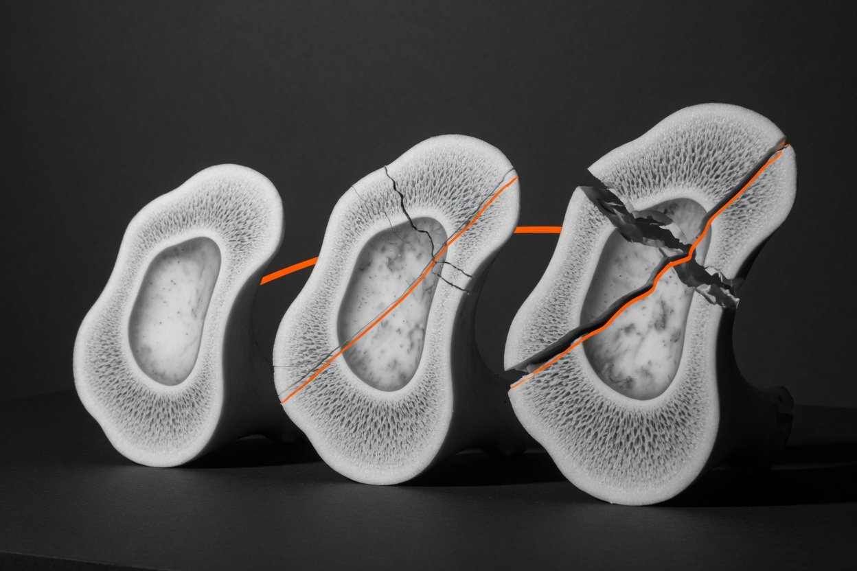
The spectrum generally progresses from an early stress response to a visible crack and, in severe cases, a complete fracture:
- Stress reaction: The bone is irritated and remodeling, but MRI does not show a definite crack.
- Stress fracture: A small fracture line has developed within the stressed bone.
- Complete fracture: The fracture extends through the bone and may affect its stability or alignment.
Not every stress reaction progresses to a fracture. Recognizing the injury early gives the bone an opportunity to recover before the damage becomes more advanced.
What does a stress reaction look like on MRI?
MRI is sensitive to changes in water content within bone and nearby soft tissues. In an early bone stress injury, a radiologist may describe bone marrow edema-type signal. This means the marrow looks brighter than expected on fluid-sensitive MRI images, suggesting that the bone is responding to excess stress.
The report may also mention periosteal edema. The periosteum is the thin tissue covering the outer surface of a bone. Edema around it is another sign of irritation caused by repetitive loading.
For example, a hip MRI may show marrow edema in the lesser trochanter region of the upper femur, along with nearby periosteal edema. If no crack is visible, the radiologist may conclude that the findings are most consistent with a stress reaction.
“No discrete fracture line is identified.”
This sentence is deliberate. It does not mean that the MRI is normal. It means there is imaging evidence of bone stress, but the radiologist cannot see a definite break in the bone.
How is a stress fracture different?
A stress fracture includes a visible fracture line. On MRI, this may appear as a thin, dark line running through part of the bone, often surrounded by marrow edema. Depending on the location and severity, the line may involve the outer cortex, the dense shell that gives bone much of its strength.
The amount of edema alone does not always determine whether an injury is called a fracture. A bone can have substantial edema without a visible crack. Conversely, a subtle fracture line may be more important for treatment planning than the overall size of the swollen area.
Radiologists therefore consider several features together:
- Whether edema is limited to the periosteum or extends into the marrow
- How much of the bone is involved
- Whether the cortex appears disrupted
- Whether a distinct fracture line is present
- The bone involved and the side of the bone under stress
Why the distinction affects activity restrictions
A stress reaction is earlier on the injury spectrum, so it may require less recovery time than a stress fracture in the same location. However, location matters as much as the label. Some bone stress injuries occur in areas that tolerate loading poorly or have a greater risk of progressing.
A clinician may recommend reducing impact activity, changing training, or temporarily limiting weight-bearing. A visible fracture line, pain during ordinary walking, or an injury in a higher-risk location may lead to stricter restrictions and closer follow-up.
Recovery cannot be predicted from one phrase in an MRI report. It depends on the specific bone, the MRI grade, symptoms, overall bone health, and whether everyday activity continues to provoke pain. Return to running or sport is usually gradual rather than based only on a fixed number of days or weeks.
What does borderline report language mean?
Imaging reports often use measured language such as compatible with, favored to represent, or no discrete fracture line identified. These phrases reflect what can and cannot be established from the images.
When a report says “stress reaction” and specifically notes the absence of a fracture line, it is placing the finding toward the earlier end of the bone stress spectrum. The wording also helps distinguish the finding from a true stress fracture, even though both injuries arise from the same basic process.
Other conditions can sometimes cause marrow edema, including a direct impact, inflammation, infection, arthritis, or a nearby tendon injury. The radiologist uses the location and pattern of the signal, along with the clinical history, to identify the most likely explanation.
Common questions about MRI findings
Is marrow edema the same as a fracture?
No. Marrow edema is a sign of increased fluid and tissue response inside the bone. It can accompany a fracture, but it can also occur in a stress reaction without a visible crack.
Can an X-ray be normal?
Yes. Early bone stress injuries may not be visible on an X-ray. MRI can detect marrow and periosteal changes before a fracture line becomes apparent on other imaging.
Does “no discrete fracture line” mean I can keep running?
Not necessarily. A stress reaction is still a bone injury, and continued painful loading may allow it to progress. Activity decisions should account for the injury’s location, symptoms, training demands, and clinical examination rather than the report wording alone.
Will another MRI be needed?
Follow-up imaging is not required in every case. Clinicians may instead monitor pain, walking tolerance, strength, and gradual return to activity. Repeat imaging may be considered when symptoms do not improve, the diagnosis remains uncertain, or the affected site carries greater risk.
When to talk to your doctor
Discuss an MRI-described stress reaction or stress fracture with a healthcare professional before returning to impact exercise. Seek prompt assessment for worsening pain, pain at rest or at night, difficulty bearing weight, sudden severe pain, or loss of normal function. This article provides general education and is not a diagnosis or a substitute for individualized medical care.
Get AI-powered analysis of your CT or MRI scan
Upload your DICOM files and receive a clear, patient-friendly report in minutes.
Analyze my scan