Lumbar Spinal Stenosis and Disc Degeneration on MRI
Learn what lumbar MRI terms like disc bulge, facet arthritis, foraminal narrowing, and spinal stenosis may mean.

Have your own scan or report? Get a clear, plain-language explanation in minutes.
General education: This article explains common MRI wording in plain language. It is not a diagnosis and cannot say what is causing any one person’s symptoms.
Why lower lumbar MRI reports often mention L4-L5 and L5-S1
The lower back carries much of the body’s weight and moves every time you bend, lift, sit, or walk. For that reason, MRI reports often describe wear-and-tear changes in the lowest lumbar levels, especially L4-L5 and L5-S1. These levels sit near the base of the spine and commonly show disc thinning, disc bulging, small bone spurs, and arthritis in the small joints at the back of the spine.
Radiology reports may use terms such as degenerative disc disease, spondylosis, facet arthropathy, foraminal stenosis, or central canal stenosis. These phrases can sound alarming, but they are descriptions of anatomy on the scan. The important question is how those findings fit with symptoms such as back pain, sciatica, numbness, leg weakness, or trouble walking.
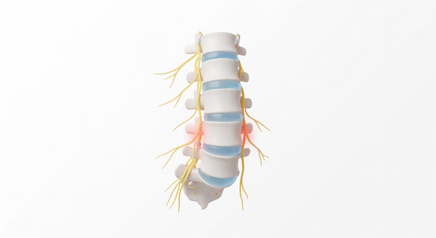
The main parts involved: discs, joints, ligaments, and nerves
Think of the lumbar spine as a stack of bones with cushions, joints, and nerve tunnels.
- Discs sit between the vertebrae and act like cushions. With degeneration, a disc can lose water, become darker on T2 MRI images, flatten, or bulge backward.
- Facet joints are small joints at the back of the spine. Arthritis can make them enlarged or irregular. Reports may call this facet arthropathy or facet hypertrophy.
- Ligamentum flavum is a ligament along the back of the spinal canal. With age or stress, it can thicken and take up more room.
- Nerves travel through the central spinal canal, pass through side zones called lateral recesses, and exit through openings called foramina.
When a bulging disc, enlarged facet joints, thickened ligaments, and bone spurs happen together, they can crowd the spaces where nerves travel. This is the basic idea behind lumbar spinal stenosis.
What spinal stenosis means
Spinal stenosis means narrowing of a nerve space. In the lumbar spine, reports usually describe three main areas:
Central canal stenosis
The central canal is the main tunnel that holds the nerve sac, also called the thecal sac or dural sac. At lower lumbar levels, this sac contains the cauda equina, a bundle of nerve roots. If the canal becomes tight, the report may say there is mild, moderate, moderate-to-severe, or severe central canal stenosis. Severe stenosis may be described as crowding of the nerve roots.
Lateral recess narrowing
The lateral recess is a side passage inside the canal where a nerve root travels before it exits. Narrowing here can irritate a traversing nerve root. For example, at L4-L5, the L5 nerve root commonly passes through this area.
Foraminal narrowing
The foramen is the doorway where a nerve exits the spine. Foraminal narrowing can be caused by disc height loss, a disc-osteophyte complex, facet arthritis, or bone spurs. At L5-S1, foraminal narrowing may affect the exiting L5 nerve root, depending on the side and severity.
How these findings can relate to symptoms
Degenerative changes can cause symptoms in different ways. Some people have MRI findings with little pain. Others have leg symptoms that closely match a compressed or irritated nerve. MRI findings are only one part of the picture.
Possible symptom patterns include:
- Low back pain: Disc degeneration, facet arthritis, muscle strain, and other causes can all contribute.
- Sciatica or shooting leg pain: This may happen when a nerve root is irritated or compressed, often from lateral recess or foraminal narrowing.
- Numbness or tingling: Nerve irritation can cause altered feeling in the buttock, thigh, calf, or foot.
- Walking limitation: Central canal stenosis can sometimes cause leg heaviness, cramping, numbness, or fatigue with standing or walking that improves with sitting or bending forward. This pattern is often called neurogenic claudication.
- Weakness: True muscle weakness can occur when nerve function is affected, but it needs clinical assessment because there are many possible causes.
Because pain can come from several structures, the exact wording on the MRI does not always predict how a person feels. A report that says mild narrowing may still matter if it matches the symptoms, while severe-looking arthritis may be painless in someone else.
Disc bulge, protrusion, and disc-osteophyte complex: what is the difference?
A disc bulge usually means the disc extends beyond its usual border over a broad area. A protrusion is a more focal extension of disc material. A disc-osteophyte complex means disc bulging is mixed with bony overgrowth, often from long-standing degeneration.
These terms do not automatically mean surgery is needed. They help explain what is taking up space. A small broad bulge may only lightly touch the nerve sac. A larger bulge combined with facet enlargement and ligament thickening can create more significant stenosis.
Why sagittal and axial MRI images may describe narrowing differently
Many people notice that one part of an MRI report says the canal does not look severely narrowed, while another part mentions moderate or severe stenosis. This can happen because MRI images look at the spine from different directions.
- Sagittal images are side-view slices. They are useful for seeing alignment, disc height, vertebral body height, and the overall shape of the spinal canal from top to bottom.
- Axial images are cross-sectional slices, like looking at a single level from above. They are especially helpful for judging the exact shape of the canal, lateral recesses, facet joints, and nerve root crowding.
A sagittal image may show only a mild or moderate front-to-back indentation of the nerve sac. But the axial image at the same level may show that the canal is tight in a cloverleaf shape because the disc bulges from the front, facet joints push in from the sides, and the ligament thickens from the back. In that situation, the axial images may describe more significant stenosis.
The reverse can also happen. A sagittal image may suggest narrowing in the side nerve opening, but axial or angled images may be needed to grade the foramen and identify which nerve, if any, is affected.
Why the report may say level numbering is approximate
Some reports mention that level numbering is approximate, especially when only one series of images is being reviewed. The radiologist usually confirms levels by counting from known landmarks on the full MRI study. If only axial images are available, it may be harder to say with complete certainty whether the tight level is L3-L4, L4-L5, or L5-S1.
This is one reason the final impression may say that findings should be correlated with the complete MRI sequences. It does not necessarily mean something is wrong with the scan; it means the full set of images gives the safest interpretation.
What mild, moderate, and severe stenosis generally suggest
There is no single phrase that perfectly predicts symptoms, but severity words give a rough sense of how much room remains around the nerves.
- Mild stenosis means there is narrowing, but the nerve space is still relatively open.
- Moderate stenosis means the space is clearly reduced and nerve contact or crowding may be possible.
- Severe stenosis means the canal or nerve opening is very tight, often with clear crowding or compression of nerve structures on imaging.
These categories should be interpreted alongside the physical exam, symptom location, walking tolerance, strength, reflexes, and medical history.
Common reassuring phrases in lumbar MRI reports
Many reports also include reassuring findings. For example, they may say there is no obvious acute fracture, no destructive bone lesion, no intraspinal mass, or no high-grade central canal stenosis on a particular sequence. These statements are helpful because they separate common degenerative changes from more urgent or less common problems.
Sometimes the report notes incidental findings, such as small kidney cyst-like areas seen at the edge of the spine images. Lumbar MRI is not designed to fully evaluate the kidneys, so these comments may be followed up only if the treating clinician thinks it is needed.
Putting the MRI into context
An MRI is a detailed map, but it is not the whole story. A useful discussion with a clinician often connects three things: where symptoms are felt, what the exam shows, and which MRI findings match that pattern. For example, pain running down the outside of the leg may be considered differently from pain centered only in the low back.
Conservative treatments, injections, and surgical options depend on the overall situation, not just one line in a report. The same MRI phrase can lead to different next steps in different people.
When to talk to your doctor
Talk with your doctor or a spine specialist if your MRI mentions moderate or severe stenosis, nerve root compression, cauda equina crowding, or foraminal narrowing that may match leg pain, numbness, weakness, or walking limitation. Also seek prompt medical attention for new loss of bladder or bowel control, numbness in the groin or saddle area, rapidly worsening weakness, fever with severe back pain, or major trauma. This article is general education and is not a personal diagnosis or treatment plan.
Get AI-powered analysis of your CT or MRI scan
Upload your DICOM files and receive a clear, patient-friendly report in minutes.
Analyze my scan