
Hip Osteonecrosis on MRI: Early Changes vs Collapse
Learn how MRI findings differ between possible early hip osteonecrosis and advanced femoral-head collapse—and why both hips are often checked.

Have your own scan or report? Get a clear, plain-language explanation in minutes.
What does hip osteonecrosis mean?
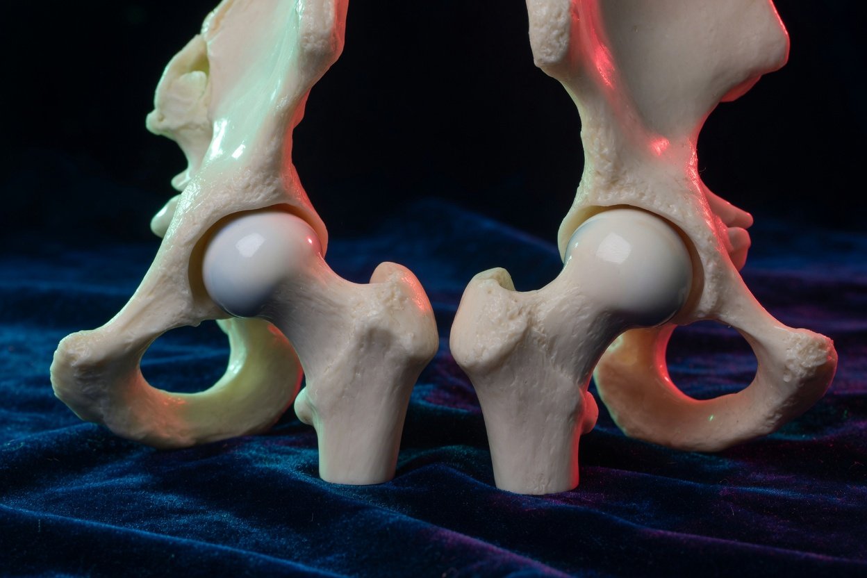
Hip osteonecrosis, also called avascular necrosis, means that part of the femoral head—the ball at the top of the thighbone—has been damaged after losing an adequate blood supply. Bone is living tissue. When its blood supply is reduced, the affected area may weaken over time.
Osteonecrosis can remain small and stable, but it can also progress. If weakened bone beneath the joint surface can no longer support normal forces, the rounded femoral head may flatten or collapse. This can damage the cartilage and lead to secondary arthritis.
Possible risk factors include a previous hip injury, prolonged or high-dose corticosteroid exposure, heavy alcohol use, certain blood disorders, autoimmune disease, and some medical treatments. In other cases, no clear cause is found.
An MRI phrase suggesting osteonecrosis does not, by itself, show how advanced the condition is. The most important distinction is whether the femoral head still has its normal shape or has begun to collapse.
What early osteonecrosis may look like on MRI
MRI can detect changes inside bone before they become visible on an X-ray. A typical early finding is a curved or serpiginous line within the femoral head. Serpiginous simply means winding or snake-like. It describes the border between affected and more normal marrow.
Radiologists look for a consistent pattern across several MRI sequences. Possible early findings include:
- A geographic or serpiginous marrow abnormality beneath the joint surface
- A low-signal line outlining part of the affected bone
- Changes on fluid-sensitive images that support an active bone response
- Preservation of the femoral head's normal rounded contour
- No definite subchondral fracture or collapse
A subtle signal change seen on only one sequence may be described as equivocal or unconfirmed. This is not the same as a definite diagnosis. Image noise, uneven fat suppression, positioning, and normal marrow variation can sometimes resemble disease. Confidence increases when the finding appears in the expected location and is reproduced on multiple types of images.
What edema and joint fluid mean
Bone marrow edema is increased fluid-related signal inside the bone. It can accompany osteonecrosis, particularly when the hip is under mechanical stress, but it is not specific to osteonecrosis. Edema can also occur with arthritis, injury, stress reaction, fracture, inflammation, or infection.
A hip joint effusion means there is extra fluid in the joint. Small effusions can be nonspecific and may occur in people without major symptoms. A larger effusion, especially with surrounding inflammation, may reflect irritation from damaged bone or cartilage. Neither edema nor joint fluid alone establishes the cause.
How collapse differs from early disease
Collapse is a structural change rather than only a marrow-signal change. MRI or X-rays may show flattening, loss of the smooth rounded contour, an irregular joint surface, or a step-like depression in the weight-bearing portion of the femoral head.
Features of more advanced disease can include:
- Subchondral fracture: a crack in weakened bone just beneath the joint cartilage
- Flattening or collapse: loss of the femoral head's normal spherical shape
- Extensive reactive edema: fluid-related change extending into the femoral neck or nearby bone
- Secondary arthritis: cartilage loss, joint-space narrowing, bone spurs, and changes in the hip socket
- Synovitis and effusion: inflammation and increased fluid within the joint
A report may say that a subchondral fracture “cannot be excluded.” That wording signals uncertainty, not confirmation. Dedicated X-rays show the hip while bearing weight, while CT can sometimes define the bone surface and a suspected fracture more clearly. The appropriate imaging depends on what is already known and whether the result would change management.
Why both hips may be reviewed
Osteonecrosis can affect both femoral heads, even when pain is present on only one side. One hip may be collapsed while the other has a smaller or earlier abnormality. For this reason, pelvic or thigh MRI examinations often include comparison views of both hips.
Comparison also helps radiologists decide whether a subtle finding is truly abnormal. If an abnormality is only partly seen or not consistently shown, a dedicated MRI of the other hip may be considered. Reviewing both sides does not mean that both hips definitely have osteonecrosis.
Common questions about an MRI report
Does “possible early osteonecrosis” mean the hip will collapse?
No. It means the images raised concern but may not have provided enough evidence for a firm diagnosis. Progression varies according to the size and location of the affected area, its cause, and whether the joint surface remains supported. Follow-up decisions require the full images, symptoms, examination, and sometimes additional imaging.
Does a lot of marrow edema mean the bone is already dead?
Not necessarily. Edema is a reactive finding rather than a direct measurement of living or dead bone. Its meaning depends on the accompanying pattern. Edema with serpiginous changes and deformity is more concerning than edema without a characteristic lesion.
Can collapse be caused by something other than osteonecrosis?
Yes. Severe osteoarthritis, a subchondral insufficiency fracture, prior trauma, infection, and less common destructive joint disorders can also deform the femoral head. Radiologists use the distribution of marrow changes, joint findings, medical history, and other studies to narrow the possibilities.
Why might an orthopedic specialist request X-rays after MRI?
MRI is highly sensitive to marrow changes, but standing or weight-bearing X-rays can show alignment, joint-space loss, and the degree of deformity in a practical way. CT may be used selectively when the precise shape of a fracture or collapse needs clarification.
How stage can affect the treatment discussion
Management is not based on the words “osteonecrosis” alone. Clinicians consider symptoms, lesion size, location, underlying cause, and whether collapse or arthritis is present. Before collapse, options may focus on monitoring, reducing stress on the joint, addressing contributing medical factors, or considering joint-preserving procedures. After substantial collapse and secondary arthritis, joint-preserving options may be less suitable, and hip replacement may enter the discussion.
No single approach is right for every person. A radiologist confirms the imaging stage, while an orthopedic hip specialist connects those findings with pain, mobility, general health, and treatment goals.
When to talk to your doctor
Discuss a report mentioning suspected osteonecrosis, subchondral fracture, deformity, or collapse with the ordering clinician. Prompt assessment is especially important for worsening groin or hip pain, difficulty bearing weight, or rapidly decreasing mobility. Sudden inability to bear weight, fever, or a hot and swollen hip warrants urgent medical attention.
This article provides general education and is not a diagnosis or a substitute for review of the complete MRI by a qualified radiologist and treating clinician.
Get AI-powered analysis of your CT or MRI scan
Upload your DICOM files and receive a clear, patient-friendly report in minutes.
Analyze my scan