Enlarged Prostate on CT: What to Know
A CT may show an enlarged prostate pressing on the bladder. Learn what it can mean, which symptoms matter, and why follow-up tests may be suggested.

Have your own scan or report? Get a clear, plain-language explanation in minutes.
What does an enlarged prostate on CT mean?
Seeing the words enlarged prostate, prostatomegaly, or prostate protruding into the bladder on a CT report can be unsettling, especially if the scan was done for pain, blood in the urine, urinary symptoms, or an unrelated abdominal problem. In many cases, an enlarged prostate is related to benign prostatic hyperplasia, often called BPH. Benign means non-cancerous, and hyperplasia means extra tissue growth.
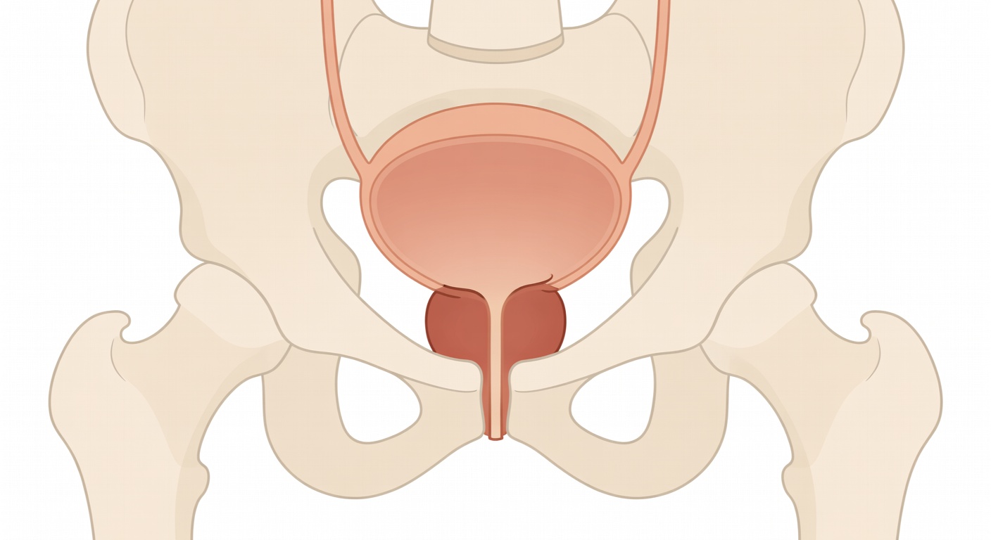
The prostate is a small gland located below the bladder and around the tube that carries urine out of the body, called the urethra. As many men age, the prostate can grow. If it grows inward or upward, it may press on the bladder outlet or push into the base of the bladder. Radiology reports may describe this as mass effect on the bladder base, intravesical protrusion, or chronic bladder outlet obstruction.
General education: This article explains common CT report language and possible next steps. It is not a diagnosis and does not replace care from your own doctor or radiologist.
Why CT scans mention the prostate and bladder
A CT scan creates cross-sectional images of the abdomen and pelvis. Even when the scan is ordered for another reason, the prostate, bladder, kidneys, ureters, bowel, bones, and blood vessels may be visible. That is why a CT report may mention prostate enlargement even if prostate disease was not the main question.
CT can show that the prostate is large or pressing into the bladder. It can also show related urinary tract findings, such as:
- Bladder wall thickening: the bladder wall looks thicker than expected.
- Trabeculation: the bladder wall appears ridged or muscular, sometimes from long-term effort to push urine past a blockage.
- Bladder distention: the bladder is very full, which may suggest trouble emptying.
- Hydronephrosis: swelling of the kidneys from backed-up urine, which is more concerning if present.
- No hydronephrosis: a reassuring phrase meaning the kidneys do not look swollen on the scan.
It is important to know that bladder wall thickening on CT is not always due to obstruction. A bladder that is not very full can look falsely thickened. Infection, inflammation, prior procedures, radiation, and other conditions can also affect the bladder wall.
Common symptoms of benign prostate enlargement
An enlarged prostate may or may not cause symptoms. When symptoms happen, they are often called lower urinary tract symptoms, or LUTS. These can include:
- Needing to urinate often, especially at night
- A sudden urge to urinate
- A weak or slow urine stream
- Starting and stopping during urination
- Straining to begin urinating
- Feeling that the bladder does not empty completely
- Dribbling after urination
- Episodes of urinary retention, meaning being unable to pass urine
Symptoms do not always match prostate size. A moderately enlarged prostate can cause major symptoms in one person, while a very large prostate may cause few symptoms in another. The exact shape of the prostate, how much it narrows the urethra, bladder muscle function, medications, infections, and neurologic conditions can all play a role.
Does an enlarged prostate on CT mean cancer?
Most prostate enlargement is benign, but CT alone cannot reliably tell the difference between BPH, prostatitis, and prostate cancer. CT is not the main test used to diagnose prostate cancer. It is also not the best test for looking inside the prostate gland in detail.
That is why a report may say that prostate enlargement is likely benign but still recommend correlation with symptoms, PSA, or urology evaluation. This does not automatically mean cancer is present. It means the CT finding should be interpreted with the whole clinical picture.
Sometimes CT reports also mention suspicious bone findings, enlarged lymph nodes, or other changes. In men, certain patterns of bone change can prompt doctors to consider prostate cancer among other possibilities. If a report mentions suspicious bone lesions or possible metastases, that is a separate and more urgent issue to review promptly with a clinician. Further testing may be needed to understand what those findings mean.
How CT differs from prostate-specific tests
CT is helpful for many abdominal and pelvic questions, such as stones, trauma, infection, bowel disease, kidney swelling, masses, or bleeding. But for the prostate, other tests often provide more useful information.
PSA blood test
PSA, or prostate-specific antigen, is a protein made by prostate tissue. PSA can rise with BPH, inflammation, infection, recent urinary retention, procedures, and prostate cancer. A PSA result is not a diagnosis by itself, but it can help guide whether more evaluation is needed.
Urinalysis and urine culture
A urinalysis checks for blood, infection markers, protein, crystals, and other clues in the urine. If infection is suspected, a urine culture may be used to identify bacteria. These tests are commonly mentioned when a CT was done for pain, urinary burning, fever, or blood in the urine.
Ultrasound and post-void residual
An ultrasound can look at the kidneys and bladder without radiation. It may measure how much urine remains in the bladder after urinating, called the post-void residual. A high residual can support the idea that the bladder is not emptying well.
Uroflowmetry and urology exam
Uroflowmetry measures how fast urine flows. A urologist may also perform a prostate exam, review medications, assess symptom severity, and decide whether medication, monitoring, or procedures should be discussed.
MRI, cystoscopy, or biopsy
In selected situations, a prostate MRI may be used to evaluate areas of concern within the prostate. A cystoscopy lets a urologist look inside the bladder and urethra with a small camera, especially when there is unexplained blood in the urine or concern for bladder problems. A biopsy is only considered when clinical findings suggest it may be needed.
What chronic outlet obstruction means
The phrase chronic bladder outlet obstruction means the bladder may have been working against resistance over time. The most common reason in older men is BPH, but it is not the only possible cause. Over time, the bladder muscle can become thicker or more ridged, which may be described as trabeculation.
If obstruction is significant, it may lead to bothersome urinary symptoms, urinary retention, bladder stones, recurrent urinary infections, or in some cases pressure backing up toward the kidneys. CT may show clues, but symptoms, urine tests, kidney function, and ultrasound findings often help determine how important the obstruction is.
Why follow-up depends on the whole picture
A CT report is one piece of information. The next step depends on why the scan was done, what symptoms are present, prior PSA results, age, medical history, medications, and the rest of the imaging findings. For example, painless blood in the urine may lead to a different workup than slow urine stream alone. A report that mentions kidney swelling, urinary retention, or suspicious bone changes may need faster attention than a report describing mild prostate enlargement only.
It can be helpful to ask the treating clinician what the prostate finding means in context, whether the bladder is emptying well, whether PSA or urine testing is appropriate, and whether urology follow-up is recommended.
When to talk to your doctor
Talk with a healthcare professional if your CT report mentions an enlarged prostate, bladder wall thickening, trabeculation, urinary blockage, hydronephrosis, blood in the urine, or suspicious bone findings. Seek prompt medical care for inability to urinate, fever with urinary symptoms, severe pelvic or flank pain, visible blood clots in the urine, new leg weakness, or rapidly worsening symptoms. This information is general education and is not a diagnosis or personal medical advice.
Get AI-powered analysis of your CT or MRI scan
Upload your DICOM files and receive a clear, patient-friendly report in minutes.
Analyze my scan