Aortic calcification on CT: an early heart-risk clue
Aortic or artery calcification on a CT report is usually not an emergency, but it can be a useful clue about long-term heart risk.

Have your own scan or report? Get a clear, plain-language explanation in minutes.
What does aortic calcification on a CT report mean?
If you had a CT scan for belly pain, chest symptoms, kidney stones, an injury, or another concern, you may be surprised to see words like aortic calcification, iliac artery calcification, coronary calcification, or vascular calcifications in the report. These findings are often listed as incidental, meaning they were seen on the scan but were not the main reason the test was ordered.
In plain language, calcification means calcium deposits are visible in the wall of an artery. In many adults, especially as people get older, this is a sign of atherosclerosis, the process sometimes called hardening of the arteries. It is usually a chronic finding that develops over time, not something that suddenly appeared on the day of the scan.
General education note: This article explains common CT wording and prevention topics. It is not a diagnosis and cannot tell you what your individual scan means.
How calcified plaque relates to heart and blood vessel risk
Arteries carry blood from the heart to the body. Over years, cholesterol-rich plaque can build up inside or along the artery wall. The body may later deposit calcium in some of these plaques. On CT, calcium is bright and easy to see, so radiologists often mention it even when the scan was done for a different reason.
Calcified plaque can be found in different places:
- Aorta: the large artery that leaves the heart and travels through the chest and abdomen.
- Iliac arteries: arteries in the pelvis that carry blood toward the legs.
- Coronary arteries: arteries that supply the heart muscle.
- Other vascular areas: such as arteries in the neck, kidneys, or legs, depending on the scan.
The important point is that calcification is a risk clue. It suggests that atherosclerosis is present somewhere in the blood vessel system. It does not automatically mean a person is having a heart attack, stroke, or blocked artery at that moment.
Why this is usually not an emergency finding
Many CT reports describe vascular calcifications as chronic, mild, moderate, scattered, or atherosclerotic. These words usually point to a long-term process. If the radiologist saw an emergency problem, the report would typically use different language, such as concern for a ruptured aneurysm, acute dissection, blocked artery, active bleeding, or another urgent diagnosis.
Calcification alone does not show whether blood flow is severely narrowed. A standard CT of the abdomen, pelvis, or chest may not be designed to measure artery blockage. It may simply show that calcium is present in the vessel wall.
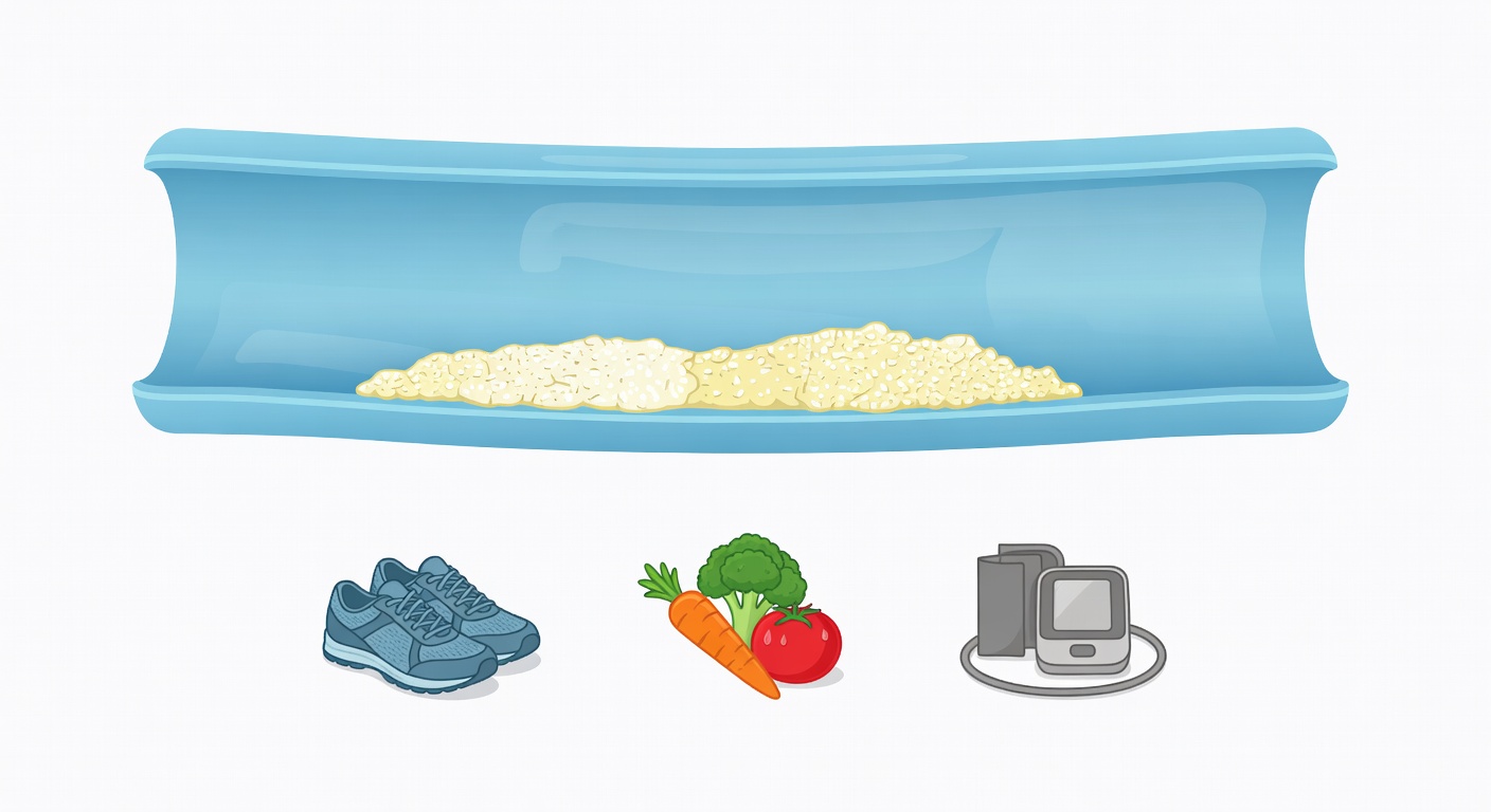
That said, incidental calcification is still worth discussing. It can be an early opportunity to review prevention: blood pressure, cholesterol, blood sugar, smoking, exercise, diet, weight, sleep, and family history.
Is aortic calcification the same as coronary artery disease?
Not exactly. Aortic calcification means plaque-related calcium is seen in the aorta. Coronary artery calcification means calcium is seen in the arteries of the heart. Both can be signs of atherosclerosis, but they are not identical findings.
When coronary calcium is seen on a regular chest CT, it may suggest increased cardiovascular risk. However, a routine CT is not always the same as a dedicated coronary artery calcium score scan. A calcium score is a specific, standardized test used in selected people to help estimate future heart risk and guide prevention decisions. Whether that test is useful depends on age, symptoms, risk factors, and what your clinician is trying to decide.
What affects whether calcifications are concerning?
The meaning of artery calcification depends on the whole clinical picture, not just one phrase in a report. Your clinician may consider:
- Age: calcification becomes more common with aging.
- Symptoms: chest pressure, shortness of breath with exertion, leg pain while walking, or stroke-like symptoms need medical attention.
- Blood pressure: high blood pressure can damage artery walls over time.
- Cholesterol levels: LDL cholesterol and other lipid measures help estimate risk.
- Diabetes or prediabetes: blood sugar problems raise vascular risk.
- Smoking history: smoking is a major risk factor for atherosclerosis.
- Kidney disease: kidney problems can be linked with more vascular calcification.
- Family history: early heart disease in close relatives can matter.
A report that says no aneurysm, no acute bleeding, or no dissection can be reassuring about emergencies, but it does not replace a cardiovascular risk review.
Questions to ask your clinician
If your CT mentions aortic, iliac, coronary, or vascular calcifications, consider bringing the report to your primary care clinician or cardiology clinician. Helpful questions include:
- Does this finding change my overall heart and stroke risk assessment?
- What are my recent blood pressure readings, cholesterol results, and blood sugar results?
- Do I need lifestyle changes, medication, or closer follow-up based on my risk profile?
- Would a coronary artery calcium score or other heart test add useful information for me?
- Are there signs of aneurysm or artery narrowing on this CT, or was it simply calcification?
These questions can turn an incidental CT finding into a prevention-focused conversation.
Prevention steps often discussed
There is no one-size-fits-all plan, and treatment decisions should be made with a clinician. In general, cardiovascular prevention often focuses on:
- Blood pressure control: checking readings and treating high blood pressure when needed.
- Cholesterol management: reviewing LDL cholesterol and whether diet, exercise, or medication is appropriate.
- Diabetes care: monitoring A1C or blood glucose if relevant.
- Not smoking: asking for support with quitting if you smoke or vape nicotine.
- Regular movement: choosing safe physical activity that fits your health status.
- Heart-healthy eating: emphasizing vegetables, fruits, whole grains, beans, nuts, fish or other lean proteins, and limiting highly processed foods.
- Weight, sleep, and stress: addressing these when they contribute to risk.
For some people, clinicians may discuss medicines such as statins, blood pressure medicines, or diabetes medicines. For others, the main focus may be tracking risk factors and making sustainable lifestyle changes.
What CT cannot tell from calcification alone
A phrase like atherosclerotic calcification does not answer every question. It may not tell:
- whether a plaque is causing a major blockage;
- whether a person needs a procedure;
- how fast the plaque is changing;
- whether symptoms are heart-related;
- which medication, if any, is right for that person.
That is why the finding should be interpreted with symptoms, examination, lab results, and the full radiology report.
When to talk to your doctor
Talk to your clinician at a routine visit if your CT report mentions aortic, iliac, coronary, or vascular calcifications, especially if you have high blood pressure, high cholesterol, diabetes, kidney disease, a smoking history, or a family history of early heart disease.
Seek urgent medical care for new or severe chest pain, trouble breathing, fainting, sudden weakness or numbness, trouble speaking, severe abdominal or back pain, or rapidly worsening symptoms. This article is general education and is not a diagnosis or individual medical advice.
Get AI-powered analysis of your CT or MRI scan
Upload your DICOM files and receive a clear, patient-friendly report in minutes.
Analyze my scan