Understanding CT Scans: A Guide for Patients
If you have been scheduled for a Computed Tomography (CT) scan, it is natural to have questions about how the technology works, its safety, and how your results are interpreted. As medical imaging evolves, particularly with the integration of Artificial Intelligence (AI), the process is becoming faster and more accurate than ever before. This guide provides a clear overview of the procedure to help you feel informed and comfortable.
What is a CT Scan?
A CT scan, also known as a CAT scan, is a diagnostic imaging procedure that uses X-rays and computer processing to create cross-sectional images (slices) of the body. While a traditional X-ray is like looking at the front of a loaf of bread, a CT scan allows doctors to look at every individual slice. This provides a much higher level of detail for bones, blood vessels, and soft tissues.
How it works:
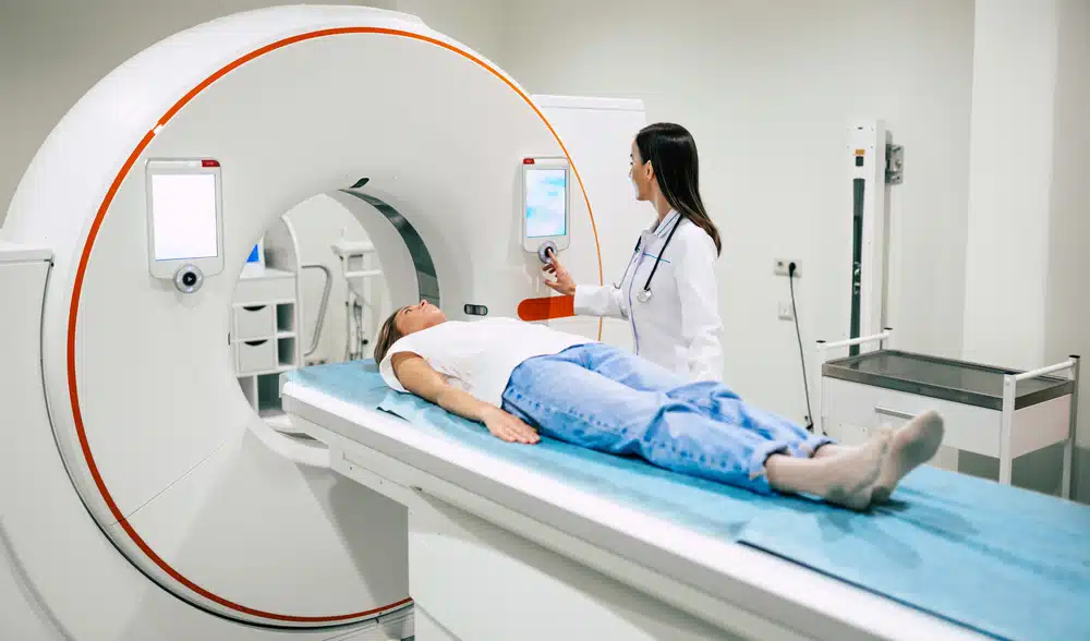
- The CT scanner is a large, doughnut-shaped machine.
- The X-ray tube rotates rapidly around your body as you lie on a motorized table.
- Detectors capture the X-ray beams and send the data to a computer, which assembles them into a detailed 3D map of your internal anatomy.
Radiation Dose: Is it Safe?
Because CT scans use X-rays, they do involve exposure to ionizing radiation. It is important to put this into perspective:
Low Risk
The amount of radiation in a single CT scan is generally small. For many patients, the diagnostic benefit of identifying a serious condition far outweighs the minimal risk associated with the radiation.
ALARA Principle
Radiologists follow a principle called ALARA (As Low As Reasonably Achievable). This means they use the lowest radiation dose possible to get the necessary image quality.
Modern Advances
Modern scanners use "dose modulation" technology to adjust the X-ray intensity based on the thickness of the body part being scanned, significantly reducing unnecessary exposure.
Contrast CT Scans and Health Effects
In some cases, your doctor may request a Contrast CT. This involves a special dye (usually iodine-based) injected into a vein or swallowed.
Purpose: Contrast acts like a "highlighter." It makes blood vessels, tumors, or inflammation stand out clearly against surrounding tissues.
What to Expect: You might feel a brief warm sensation throughout your body or a metallic taste in your mouth during the injection.
Kidney Function
Because the kidneys filter the contrast dye out of your body, your doctor may check your kidney function (via a blood test) before the scan.
Allergies
While rare, some people are allergic to iodine contrast. Be sure to tell the staff if you have had a reaction to contrast in the past or if you have asthma.
Hydration
You will likely be encouraged to drink plenty of water after the scan to help flush the dye from your system.
CT vs. MRI: What's the Difference?
Patients often wonder why they are getting a CT instead of an MRI. While both provide detailed images, they serve different purposes:
| Feature | CT Scan | MRI (Magnetic Resonance Imaging) |
|---|---|---|
| Technology | Uses X-rays (Radiation) | Uses Magnets and Radio Waves |
| Speed | Very fast (minutes) | Slower (20–60 minutes) |
| Best for... | Bone fractures, lung issues, chest/abdomen, and emergencies (trauma). | Soft tissues, ligaments, tendons, brain, and spinal cord. |
| Patient Experience | Quiet; better for claustrophobia. | Very loud; requires lying very still in a narrow tube. |
How CT Scans are Evaluated: Radiologists and AI
Once your scan is complete, the images must be analyzed. Traditionally, this is the job of a Radiologist—a specialized doctor who "reads" the images to look for abnormalities. Today, the process is being enhanced by Artificial Intelligence (AI). Here is how they work together:
1. The Role of AI
AI software can scan thousands of images in seconds. It acts as a "second pair of eyes" that never gets tired. AI is particularly good at:
- Flagging Urgent Cases: AI can instantly identify life-threatening issues (like a brain bleed or a lung clot) and move those scans to the top of the radiologist's pile.
- Precise Measurements: AI can measure the volume of a tumor or the density of a bone with mathematical precision that is difficult to achieve by eye alone.
2. The Role of the Radiologist
The radiologist remains the final authority. They use the AI's findings to provide a comprehensive diagnosis, taking into account your medical history, symptoms, and the subtle nuances of the images.
The result? A faster, more accurate diagnosis that combines human expertise with machine efficiency.