Sclerotic Bone Lesions and Prostate Cancer Imaging
Sclerotic bone lesions on CT can be worrying. Learn why PSA testing and newer imaging like PSMA PET/CT may be discussed.

Have your own scan or report? Get a clear, plain-language explanation in minutes.
What does “sclerotic bone lesion” mean on a CT report?
Seeing the words “multiple sclerotic bone lesions” or “osteoblastic metastases” on a CT report can be frightening. A sclerotic bone lesion is an area of bone that looks denser or whiter than expected on imaging. “Osteosclerotic” and “osteoblastic” are related terms that describe increased bone formation or mineral density in a spot or in many spots.
Importantly, a CT description is not the same as a confirmed diagnosis. Radiologists use patterns, medical history, lab results, and sometimes additional imaging to decide what a finding is most likely to represent. This article is general education, not a diagnosis, and it cannot replace review by your own clinician and radiologist.
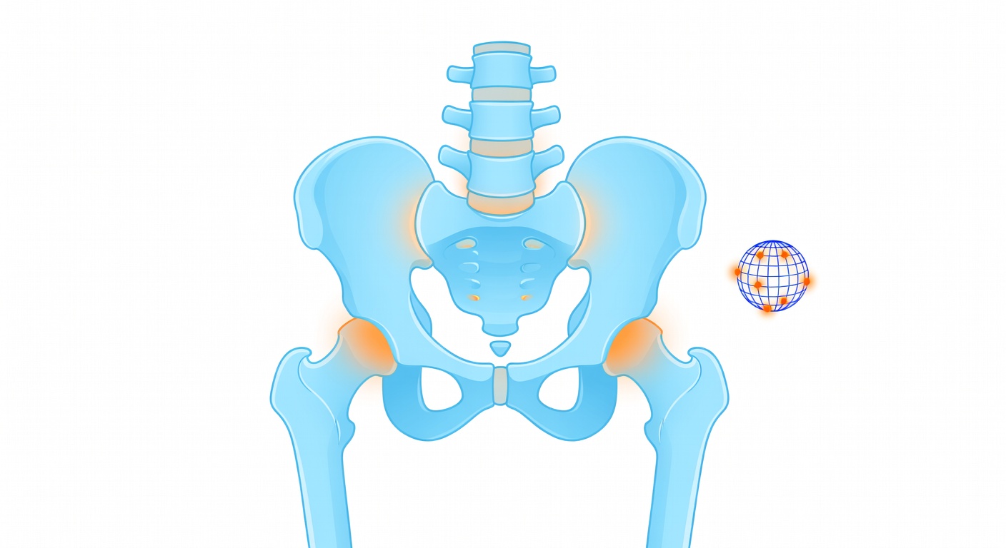
Key point: Sclerotic bone lesions have several possible causes. When they are numerous and scattered through the spine, pelvis, ribs, or hips, doctors often consider metastatic cancer among the possibilities—especially in men when the prostate is enlarged or PSA is abnormal.
Why prostate cancer is often mentioned
Prostate cancer has a particular tendency to spread to bone when it becomes advanced. When prostate cancer involves bone, it often produces osteoblastic changes, meaning the affected areas may look denser on CT or X-ray. This is different from some other cancers that more often create bone loss or “lytic” lesions.
Common places for prostate cancer bone spread include the spine, pelvis, ribs, and upper thigh bones. These are also areas often included on abdominal or pelvic CT scans. A report may say the pattern is “suspicious for osteoblastic metastases” if many small dense spots are seen in these locations.
However, an enlarged prostate does not automatically mean prostate cancer. Benign prostatic hyperplasia, often called BPH, is very common with aging and can enlarge the prostate without cancer. That is why doctors usually look at the whole picture: symptoms, exam findings, PSA level, previous imaging, and sometimes biopsy results.
What else can cause sclerotic bone lesions?
Although metastatic prostate cancer is an important consideration, it is not the only cause of sclerotic bone findings. Possible explanations may include:
- Benign bone islands, which are small dense areas that are usually harmless.
- Healing fractures or areas of old injury.
- Degenerative changes related to arthritis, especially near joints or the spine.
- Paget disease of bone, a condition that changes bone remodeling.
- Other cancers that can cause sclerotic or mixed bone changes.
- Prior treatment effects, depending on a person’s cancer history and therapies.
The pattern matters. A single stable bone island is very different from many new dense spots throughout the skeleton. This is why comparison with older scans can be extremely helpful.
Why PSA testing may be discussed
PSA stands for prostate-specific antigen. It is a blood test related to prostate tissue. PSA can rise for several reasons, including prostate enlargement, inflammation, infection, recent procedures, and prostate cancer. It is not a perfect test, but it can provide important context when imaging raises concern for prostate disease.
If a CT report mentions suspicious sclerotic bone lesions in a man, clinicians may discuss PSA testing or review prior PSA results. A very elevated PSA may increase concern for prostate cancer, while a lower PSA may shift the discussion toward other explanations. Either way, PSA is interpreted along with the rest of the medical evaluation.
How traditional bone imaging has been used
For many years, one common test for possible prostate cancer spread to bone has been a bone scan, also called bone scintigraphy. This test uses a small amount of radioactive tracer to show areas of increased bone activity. It can detect many bone metastases, but it may also light up in areas of arthritis, fractures, or other non-cancer changes.
CT and MRI can also help. CT is good at showing dense bone lesions and other anatomy, while MRI can be especially useful for evaluating the spine, bone marrow, nerves, and spinal cord if symptoms suggest a complication. The best test depends on the clinical question, the person’s history, local availability, and the treating team’s judgment.
What is PSMA PET/CT?
PSMA PET/CT is one of the major advances in prostate cancer imaging. PSMA stands for prostate-specific membrane antigen, a protein often found at higher levels on prostate cancer cells. In a PSMA PET/CT scan, a tracer that targets PSMA is injected into the bloodstream. The PET part shows areas where the tracer collects, and the CT part provides anatomical detail.
PSMA PET/CT is increasingly used to help detect and stage prostate cancer, including disease in lymph nodes and bones. It can be useful in some people with newly diagnosed higher-risk prostate cancer and in some people whose PSA rises after treatment. It may find disease that is difficult to see on conventional imaging, although it is not needed for every patient.
Like all imaging, PSMA PET/CT is not perfect. Some prostate cancers may show little PSMA uptake, and some non-prostate conditions can occasionally take up tracer. Results must be interpreted by specialists in the context of the patient’s history, PSA, pathology, and other imaging.
What a suspicious CT impression usually triggers
When a CT report describes multiple osteosclerotic lesions suspicious for metastases, the next steps are usually about confirmation and staging—not jumping to conclusions from one phrase. The clinical team may consider:
- Reviewing the full CT study, including all contrast phases and bone windows.
- Comparing with prior CT, MRI, X-ray, or bone scan images.
- Checking PSA and other blood tests, such as kidney function or alkaline phosphatase when relevant.
- Arranging urology or oncology evaluation if prostate cancer is suspected.
- Ordering additional imaging, such as PSMA PET/CT, bone scan, MRI, or targeted CT, depending on the case.
- Considering tissue diagnosis, such as prostate biopsy, if cancer has not already been confirmed and the clinical picture supports it.
Sometimes CT reports also mention other findings, such as a kidney cyst, diverticulosis, bladder wall changes, or atherosclerosis. These may be important, but they are separate from the question of whether bone lesions represent cancer spread.
Symptoms that may matter
Some people with bone metastases have pain, while others have few or no symptoms. Doctors may ask about persistent back, hip, rib, or pelvic pain; unexplained weight loss; urinary symptoms; blood in the urine; weakness; numbness; or trouble walking. They may also ask about fever, recent infection, falls, known cancers, and prior surgeries or radiation therapy.
New severe back pain with leg weakness, numbness, or loss of bladder or bowel control can be urgent because it may suggest pressure on nerves or the spinal cord. This article cannot determine whether any symptom is related to imaging findings, but symptoms help clinicians decide how quickly and what type of evaluation is needed.
Reading the wording carefully
Radiology reports often use probability language. “Suspicious for” means the imaging pattern raises concern, not that the diagnosis is proven. “Correlation with PSA” means the radiologist is recommending that the imaging be interpreted together with prostate-related blood tests and clinical history. “Consider PSMA PET/CT” means the scan may be helpful, but the ordering clinician decides based on the full situation.
If you are reading a report in translation, wording can feel even more alarming. It is reasonable to ask the doctor to explain the impression in plain language and to clarify what is known, what is uncertain, and what test is meant to answer the next question.
When to talk to your doctor
Talk with your doctor promptly if your CT report mentions multiple sclerotic bone lesions, osteoblastic metastases, or a recommendation to check PSA or obtain further bone imaging. A clinician can review the full scan, compare prior studies, order appropriate tests, and decide whether urology or oncology referral is needed.
Seek urgent medical care if you have severe or worsening bone or back pain, new leg weakness or numbness, trouble urinating, loss of bladder or bowel control, fever, blood in the urine, or sudden worsening of your general condition. This content is for general education only and is not a diagnosis or individualized medical advice.
Get AI-powered analysis of your CT or MRI scan
Upload your DICOM files and receive a clear, patient-friendly report in minutes.
Analyze my scan