
How to Read a Knee MRI After ACL or Meniscus Surgery
Postoperative knee MRI reports use a language of their own. Learn what graft signal, repair-site changes, and uncertain findings may mean.

Have your own scan or report? Get a clear, plain-language explanation in minutes.
Why postoperative knee MRI reports sound different
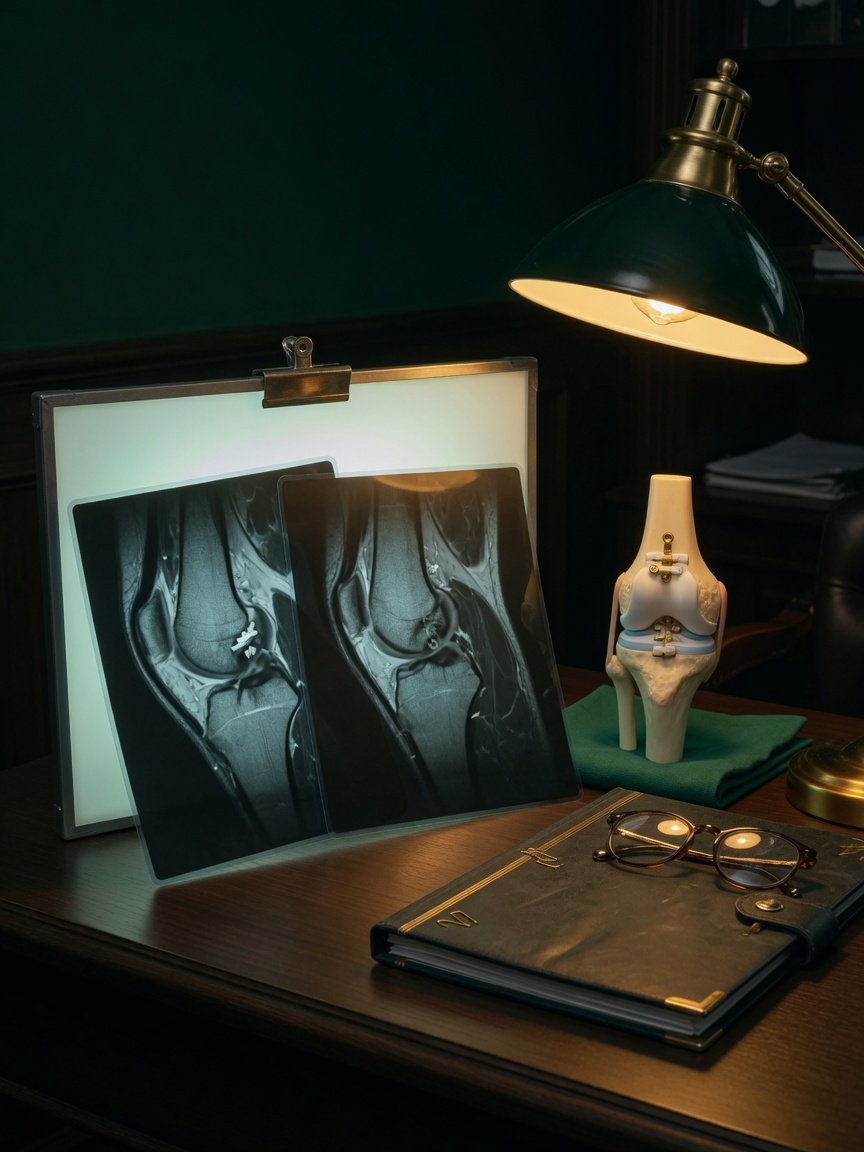
Reading a knee MRI after ACL reconstruction or meniscus surgery is not the same as reading a scan of a knee that has never been operated on. Surgery intentionally changes the knee’s anatomy. A reconstructed ligament may have a different signal from a native ligament, a repaired meniscus may retain a bright line at the repair site, and fixation devices can distort part of the image.
That is why postoperative reports often include cautious wording. The radiologist must separate expected healing and surgical changes from complications such as a graft tear or recurrent meniscus tear. Symptoms, the operative report, the physical examination, and comparison with earlier scans can all help.
After surgery, an MRI finding is only one part of the story. The most useful interpretation connects the images with the operation performed and how the knee is functioning now.
ACL reconstruction: understanding the graft vocabulary
“Intact,” “continuous,” and “normal fibrillar signal”
After ACL reconstruction, the report may refer to the reconstructed ACL as the graft, neoligament, or—in some international reports—neolegamento. These terms describe the tendon tissue placed in the knee to perform the job of the original ACL.
Reassuring descriptions include:
- Intact graft: The fibers do not show a complete disruption.
- Continuous graft: The tissue can be followed between its attachment points.
- Normal fibrillar signal: The graft has an organized, fiber-like appearance.
- No evidence of re-tear: The scan does not show imaging signs of a new rupture.
A report may also describe the graft’s direction, tension, or relationship to the roof of the notch in the femur. These details help assess whether the graft follows an expected course.
Graft signal and “ligamentization”
An ACL graft changes as it heals and remodels. This process is sometimes called ligamentization. The graft can temporarily appear brighter on certain MRI sequences than a normal native ACL. Signal within or around the graft is therefore not automatically a tear.
Earlier imaging may mention graft-site edema, meaning extra fluid-like signal near the reconstructed ligament. A later report may say that this edema has resolved while the neoligament remains continuous. Comparison with prior scans helps the radiologist recognize this progression.
Fixation anchors and metal artifact
ACL grafts are secured in bone tunnels with devices such as screws, buttons, or anchors. Some devices produce susceptibility artifact: a local blur, dark area, or distortion caused by metal interacting with the MRI magnet.
Artifact at a known fixation site is often an expected postoperative finding. It does not by itself mean the device is loose or the graft has failed. However, artifact can hide nearby structures, so the report may state that assessment is limited in that area.
Meniscus repair: why the report may not give a yes-or-no answer
A repaired meniscus can be especially difficult to evaluate. Before surgery, abnormal signal reaching the meniscus surface may support a diagnosis of a tear. After repair, that same region can remain altered because of scar tissue, healing tissue, sutures, or residual changes from the original tear.
A common report phrase is:
“Altered undersurface signal, possibly postoperative in nature, although a component of tearing is not excluded.”
This wording does not confirm a re-tear. It means the MRI appearance overlaps with both postoperative change and tearing, so the images alone cannot reliably settle the question. The phrase “not excluded” describes uncertainty rather than a definite diagnosis.
Radiologists may specifically request the operative history because it matters whether the meniscus was repaired, partially removed, or treated in another way. Knowing the original tear’s location also helps determine whether a current signal abnormality is new or expected.
After partial meniscectomy: trimming versus another tear
A partial meniscectomy removes damaged meniscus tissue and reshapes the remaining edge. On a later MRI, the meniscus may look smaller, blunted, shortened, or irregular. These findings can represent expected surgical trimming rather than recurrent injury.
The radiologist looks for changes that extend beyond the expected surgical margin, displaced tissue, or a new pattern compared with earlier imaging. Even then, distinguishing postoperative contour from a recurrent tear may be difficult without the operative report and clinical context.
What if the MRI finds a tear but the knee has no symptoms?
Sometimes a routine or surveillance scan reports a complex meniscus tear even though the referral says there are no clinical symptoms. The ACL graft may look normal at the same time. This can feel alarming, but MRI findings and symptoms do not always match.
A newly reported finding may represent a true structural change, a longstanding abnormality that is more visible on the current scan, or a postoperative appearance interpreted differently by another reader. An imaging finding alone does not determine whether treatment is needed. Clinicians usually consider pain, swelling, locking, instability, activity level, examination findings, and comparison with previous images.
Do not overlook the rest of the knee
Postoperative MRI is not limited to the ACL or meniscus. The report may also discuss cartilage, bone marrow, other ligaments, tendons, joint fluid, or the kneecap joint. For example, a scan after meniscus surgery may show expected trimming while noting that cartilage wear behind the kneecap has progressed.
This does not necessarily mean the surgery caused the cartilage change. It means the radiologist observed a difference that may deserve clinical context, especially if there is new pain at the front of the knee, stiffness, or swelling.
A practical way to read the report
- Start with the surgical target: Is the ACL graft intact? Does the meniscus show expected repair or trimming?
- Look for comparison language: Words such as stable, improved, resolved, new, or progressed are often more informative than an isolated finding.
- Separate certainty from caution: “Torn” is different from “a component of tearing is not excluded.”
- Note technical limits: Metal artifact or postoperative distortion may reduce confidence.
- Read the impression with the full report: The final summary is useful, but the detailed findings explain why the radiologist reached that conclusion.
- Match the scan to the knee: Symptoms and examination findings help determine whether an MRI abnormality is clinically important.
When to talk to your doctor
Discuss the report with your surgeon or another qualified clinician if it mentions graft disruption, a possible recurrent tear, displaced meniscus tissue, worsening cartilage damage, or findings limited by artifact. Prompt assessment is also appropriate for new instability, repeated locking, major swelling, or difficulty bearing weight. This article provides general education only and cannot diagnose a condition or replace an individualized medical evaluation.
Get AI-powered analysis of your CT or MRI scan
Upload your DICOM files and receive a clear, patient-friendly report in minutes.
Analyze my scan