MRCP explained: bile ducts, gallstones, and pancreas findings
Learn what MRCP checks, why gallstones can be hard to see, and how pancreatic cysts or pancreatitis are interpreted with labs and symptoms.

Have your own scan or report? Get a clear, plain-language explanation in minutes.
What is MRCP?
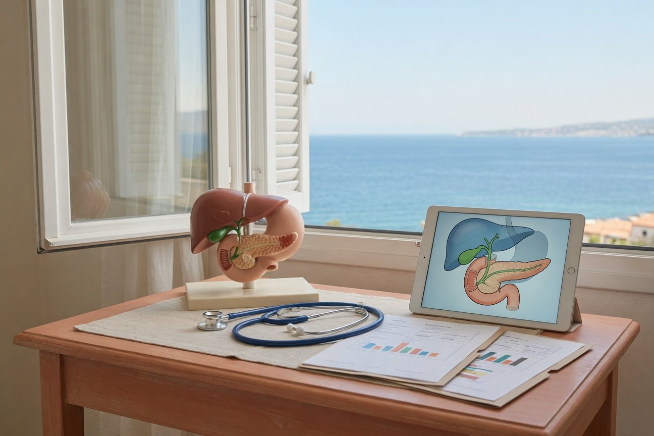
MRCP stands for magnetic resonance cholangiopancreatography. It is a special type of MRI that focuses on the fluid-filled tubes that carry bile and pancreatic juices. These include the bile ducts, the gallbladder, and the pancreatic duct.
MRCP does not use X-rays. Many MRCP exams are done without contrast dye, although some abdominal MRI studies include contrast to better evaluate the liver, pancreas, kidneys, or masses. The MRCP part uses heavily fluid-sensitive MRI pictures, so bile and pancreatic fluid appear bright while stones, narrowing, or blockages may appear as dark gaps or changes in the duct shape.
MRCP is best understood as a map of the bile and pancreatic drainage system. It is powerful, but it is not the whole story by itself.
This article is for general education only. It is not a diagnosis and cannot replace the official radiology report or a discussion with your clinician.
What does an MRCP look for?
An MRCP is commonly ordered when doctors are concerned about gallstones, bile duct blockage, pancreatitis, abnormal liver blood tests, jaundice, or an unclear finding on CT or ultrasound.
It can help evaluate:
- Gallbladder stones, also called cholelithiasis.
- Common bile duct stones, also called choledocholithiasis.
- Bile duct widening, which may suggest a blockage downstream.
- Bile duct narrowing or strictures.
- Pancreatic duct dilation or an abrupt duct cutoff.
- Inflammation around the pancreas, especially when MRI sequences beyond MRCP are included.
- Small pancreatic cysts or side-branch duct changes.
Some reports mention that the bile duct is mildly prominent but tapers smoothly. This often means the duct is a little larger than expected, but there is no obvious abrupt blockage. Whether that matters depends on age, prior gallbladder surgery, symptoms, and blood tests.
Gallstones: why one report may see them and another may not
A common patient worry is: Were my gallstones missed? The answer is that gallstones can be sequence-dependent. On MRI, stones may show up clearly on one set of images but be subtle or not visible on another. Small stones, sludge, or tiny crystals can be especially difficult to prove.
Ultrasound is often very good at seeing stones inside the gallbladder. MRCP is especially helpful for looking at the bile ducts, including whether a stone has moved from the gallbladder into the common bile duct. CT can miss some gallstones because many stones are not calcified enough to stand out.
So, it is possible for an MRCP report to say likely gallstones in the gallbladder while also saying no definite stone in the common bile duct. Those are different findings. Gallstones sitting in the gallbladder do not always mean there is an active blockage.
What MRCP can and cannot rule out
MRCP is very useful, but it has limits. A normal-looking MRCP can make a major bile duct blockage less likely, especially if the ducts are not dilated and no filling defect is seen. However, it may not rule out every small stone, passed stone, sludge, early inflammation, or intermittent blockage.
MRCP may be limited by:
- Breathing or motion blur during the scan.
- Very small stones or microlithiasis.
- Thick-slice images that blur tiny details.
- Non-contrast technique when a mass or inflammation needs more detail.
- Normal anatomy variants, bowel fluid, or duodenal diverticula that can mimic nearby cystic structures.
This is why radiology reports often use phrases such as no definite, not clearly seen, indeterminate, or correlate clinically. These words do not always mean something dangerous. They mean imaging has to be interpreted with the rest of the medical picture.
Pancreatitis on MRI and MRCP
Pancreatitis means inflammation of the pancreas. On MRI, acute interstitial or edematous pancreatitis may appear as swelling, fluid, or inflammation around the pancreas. Reports may describe edema near the pancreatic head, neck, body, or tail.
MRCP also helps check whether pancreatitis might be related to a bile duct stone. If the bile ducts and pancreatic duct are not widened and no common bile duct stone is seen, a large ongoing blockage is less likely. But small stones or sludge can still be difficult to detect, especially if a stone has already passed.
For pancreatitis, doctors usually rely on a combination of:
- Symptoms, such as upper abdominal pain, nausea, or vomiting.
- Blood tests, especially lipase or amylase.
- Liver tests and bilirubin to look for a bile-related cause.
- Imaging to look for complications, duct blockage, or another diagnosis.
An MRCP report that says no necrosis, no large fluid collection, and no duct dilation is generally describing the absence of certain visible complications. It does not, by itself, grade how sick a person feels or decide treatment.
Tiny pancreatic cysts: common words in reports
Many patients are surprised when an MRCP mentions tiny bright spots in the pancreas. These may be described as:
- Small pancreatic cystic foci.
- Side-branch duct ectasia, meaning tiny side branches of the pancreatic duct look a bit widened.
- Side-branch IPMN-type cysts, referring to a type of pancreatic cyst that can communicate with the duct system.
- Small pseudocyst, especially after pancreatitis.
- Indeterminate fluid focus, meaning the radiologist cannot fully classify it from the images alone.
Very small pancreatic cysts are often found incidentally. The key details are the size, whether the main pancreatic duct is widened, whether there is a solid component or nodule, whether the cyst is growing, and whether symptoms or pancreatitis are present.
If a report recommends follow-up, that does not automatically mean cancer is suspected. Follow-up MRI or MRCP is often used to confirm stability over time. Your doctor may also compare the scan with older CT, MRI, or ultrasound studies.
Pancreatic head, uncinate process, and duodenum: why this area can be confusing
The pancreatic head sits next to the duodenum, the first part of the small intestine. The uncinate process is a hook-like part of the pancreas that curves behind nearby blood vessels. This region is crowded: bile ducts, pancreatic ducts, bowel loops, fluid, and sometimes diverticula are close together.
A report may mention uncertainty near the pancreatic head, uncinate process, or duodenum. Sometimes a fluid-filled duodenal diverticulum, bowel contents, a small cyst, or a duct structure can look similar on limited sequences. This is one reason radiologists review all sequences together and may recommend comparison, repeat MRI, endoscopic ultrasound, or follow-up only if the finding remains unclear or concerning.
Why labs and symptoms matter as much as imaging
MRCP answers imaging questions. It does not replace the clinical picture. For bile duct and pancreas concerns, doctors often compare the scan with:
- Bilirubin, which can rise with bile blockage and jaundice.
- Alkaline phosphatase, GGT, ALT, and AST, which help evaluate liver and bile duct irritation.
- Lipase or amylase, which help assess pancreatitis.
- White blood cell count and CRP, which may support inflammation or infection.
- Fever, jaundice, pain pattern, vomiting, dark urine, and weight changes.
For example, a scan showing gallstones but normal bile ducts may be interpreted differently in someone with no symptoms than in someone with right upper abdominal pain, fever, jaundice, and abnormal liver tests.
Questions to ask after an MRCP report
If your report mentions gallstones, pancreatitis, bile duct prominence, or pancreatic cysts, consider asking your clinician:
- Are there stones in the gallbladder, the bile duct, or both?
- Are the bile ducts or pancreatic duct dilated?
- Do my bilirubin and liver enzymes suggest a current or recent blockage?
- Do my lipase or amylase levels fit with pancreatitis?
- What size are the pancreatic cysts, and are there any high-risk features?
- Should this be compared with prior imaging?
- Is follow-up MRI/MRCP, ultrasound, endoscopic ultrasound, or specialist review recommended?
When to talk to your doctor
Talk with your doctor about any MRCP result you do not understand, especially if the report mentions bile duct blockage, pancreatitis, an indeterminate pancreatic finding, or a pancreatic cyst needing follow-up.
Seek urgent medical care if you develop severe or worsening abdominal pain, fever, yellowing of the skin or eyes, dark urine, persistent vomiting, dehydration, black stools, vomiting blood, fainting, or feeling very unwell. Imaging is only one part of care, and symptoms that are new or worsening should be assessed promptly.
Get AI-powered analysis of your CT or MRI scan
Upload your DICOM files and receive a clear, patient-friendly report in minutes.
Analyze my scan