Mild Disc Bulges on MRI: What They Usually Mean
Mild disc bulges and spine wear on MRI are common. Learn how doctors match imaging findings with symptoms and exams.

Seeing a mild disc bulge on your MRI report can feel alarming
Words like disc bulge, spondylosis, facet arthritis, or degenerative disc disease can sound serious. Many people read an MRI report and worry that these findings must be the cause of every ache, twinge, or episode of pain.
In many cases, mild disc bulges and mild wear-and-tear changes are common MRI findings, especially in the neck and lower back. They may be related to symptoms, but they also may be incidental, meaning they are seen on the scan but are not the main reason a person hurts.
This article is general education, not a diagnosis. MRI results should be interpreted by a qualified clinician or radiologist together with your symptoms, physical exam, and full medical history.
What is a disc bulge?
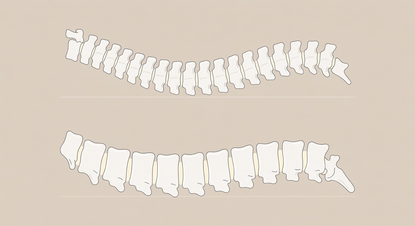
Spinal discs sit between the bones of the spine, called vertebrae. They act like cushions and help the spine bend, twist, and absorb stress. A disc has a tougher outer layer and a softer center.
A disc bulge means the outer edge of the disc extends slightly beyond its usual border. This is often broad and shallow rather than a large focal rupture. A bulge is different from a large disc herniation or extrusion, where disc material may push out more focally and potentially irritate a nearby nerve.
MRI reports may describe a bulge as:
- Mild, small, or shallow
- Broad-based, meaning it involves a wider portion of the disc edge
- Causing mild thecal sac indentation, meaning it slightly touches or presses on the lining around the nerves
- Not causing high-grade stenosis, meaning there is no severe narrowing seen
These details matter. A mild bulge that does not significantly narrow the spinal canal or nerve openings is usually interpreted very differently from a large herniation that clearly compresses a nerve root or the spinal cord.
What do spondylosis and facet arthritis mean?
Spondylosis is a general term for age-related or wear-and-tear changes in the spine. It may include disc drying, small bone spurs, mild disc space narrowing, or joint changes.
Facet joints are small joints at the back of the spine. Like knees, hips, and hands, these joints can develop arthritis over time. MRI or CT reports may call this facet arthropathy or facet arthritis.
These changes do not automatically mean something is dangerous or worsening quickly. They are part of the way many spines change over time. The important question is whether the location and severity of the findings match the person’s symptoms.
Why mild findings may not explain all pain
MRI is very sensitive. It can show small changes in discs, joints, bones, ligaments, and soft tissues. That is helpful, but it also means MRI often finds changes that are not necessarily painful.
For example, a person may have a mild lumbar disc bulge at L5-S1 but feel pain mainly in the upper back or neck. In that case, the low back finding may not explain the current pain pattern. Another person may have mild cervical disc bulges but no arm pain, numbness, weakness, or signs of spinal cord irritation. The imaging finding may be real, but not clinically important at that time.
Pain is complex. Muscles, joints, nerves, sleep, stress, activity level, prior injuries, inflammation, and general health can all influence how the back or neck feels. MRI is one piece of the puzzle, not the entire story.
How doctors match MRI findings to symptoms
Clinicians usually ask whether the MRI finding matches a recognizable pattern. This is sometimes called clinical correlation. It means the scan must be interpreted in context.
Neck findings and arm symptoms
In the cervical spine, or neck, a disc bulge or bone spur may matter more if it narrows a nerve opening and the person has symptoms along that nerve’s path. This may include pain, tingling, numbness, or weakness that travels from the neck into the shoulder, arm, or hand.
A report that says there is no spinal cord compression and no abnormal cord signal is often reassuring, but it does not replace a neurological exam. Axial MRI images and foraminal views are especially important for checking the nerve openings.
Low back findings and leg symptoms
In the lumbar spine, or lower back, disc changes at L4-L5 or L5-S1 are commonly discussed because these levels carry a lot of load. A finding may be more meaningful if symptoms travel in a pattern down the buttock, thigh, calf, or foot, especially if the exam suggests a specific nerve root is irritated.
On the other hand, mild lower lumbar disc bulging without clear nerve compression may or may not be the main cause of back pain. Many reports describe the spinal canal as open or without severe stenosis, which suggests there is no obvious high-grade central narrowing on the reviewed images.
What does it mean when the report says there is no severe compression?
Many MRI reports use phrases such as no high-grade canal stenosis, no definite cord compression, or no large disc herniation. These are generally reassuring phrases. They suggest that, on the images reviewed, there is no severe narrowing of the central spinal canal or obvious major pressure on the spinal cord or nerve bundle.
However, mild or moderate narrowing of smaller nerve openings can sometimes be harder to judge, especially if only limited image sequences are available. That is why the complete MRI exam, including sagittal and axial views, matters. It is also why the official radiology report and clinical exam are so important.
Everyday spine-health basics
For many people with mild degenerative spine findings, the conversation shifts from the MRI image to overall spine function. General spine-health habits may include:
- Movement variety: The spine usually tolerates changing positions better than staying in one posture for long periods.
- Posture flexibility: There is no single perfect posture. Sitting, standing, walking, and resting in different positions can reduce repetitive strain.
- Strength and conditioning: A stronger trunk, hips, shoulders, and legs can support everyday activities and may improve confidence with movement.
- Sleep and recovery: Poor sleep can make pain feel more intense. A consistent sleep routine and comfortable sleep setup may support recovery.
- Weight management and general health: Overall health, including activity level, nutrition, and body weight, can affect how the spine handles daily load.
- Stress awareness: Stress does not mean pain is imaginary, but it can influence muscle tension, pain sensitivity, and coping.
These are broad wellness concepts, not individualized treatment instructions. The best approach depends on the person, the symptoms, the exam, and any other health conditions.
Questions to ask about a mild disc bulge
If your report mentions mild disc bulges, facet arthritis, or spondylosis, it may help to ask your clinician:
- Does this MRI finding match my pain pattern?
- Is there any nerve root compression or spinal cord compression?
- Are the nerve openings, called foramina, narrowed?
- Were all MRI sequences reviewed, including axial images?
- Are there any findings that require urgent attention?
- What non-surgical and lifestyle options are appropriate to discuss in my situation?
These questions can shift the discussion from simply naming MRI findings to understanding what they mean for your symptoms and next steps.
When to talk to your doctor
Talk to your doctor or another qualified healthcare professional if you have back or neck pain that is persistent, worsening, or affecting daily life. Seek prompt medical care for new weakness, trouble walking, numbness in the groin or saddle area, loss of bladder or bowel control, fever with spine pain, major trauma, or rapidly worsening numbness or pain.
Remember: mild disc bulges on MRI are often wear-and-tear findings. Their importance depends on whether they match your symptoms, exam findings, and the full imaging report.
Get AI-powered analysis of your CT or MRI scan
Upload your DICOM files and receive a clear, patient-friendly report in minutes.
Analyze my scan