Knee MRI: Meniscus Tears, Effusions, and Baker Cysts
Learn what common knee MRI findings like meniscus tears, small effusions, osteoarthritis, and Baker cysts may mean.

Have your own scan or report? Get a clear, plain-language explanation in minutes.
What a knee MRI can show
A knee MRI is often ordered when someone has knee pain, swelling, catching, locking, or an injury during sports or daily activity. MRI is useful because it can show soft tissues that X-rays cannot see well, including the menisci, ligaments, cartilage, tendons, bone marrow, and joint fluid.
Many MRI reports include phrases such as possible medial meniscus tear, small joint effusion, mild osteoarthritis, Baker cyst, or possible discoid lateral meniscus. These findings can sound alarming, but their importance depends on the full MRI study, the physical exam, and the pattern of symptoms.
Important: This article is general education, not a diagnosis. MRI findings should be interpreted by a qualified clinician or radiologist in the context of your symptoms and exam.
The meniscus: the knee’s shock absorber
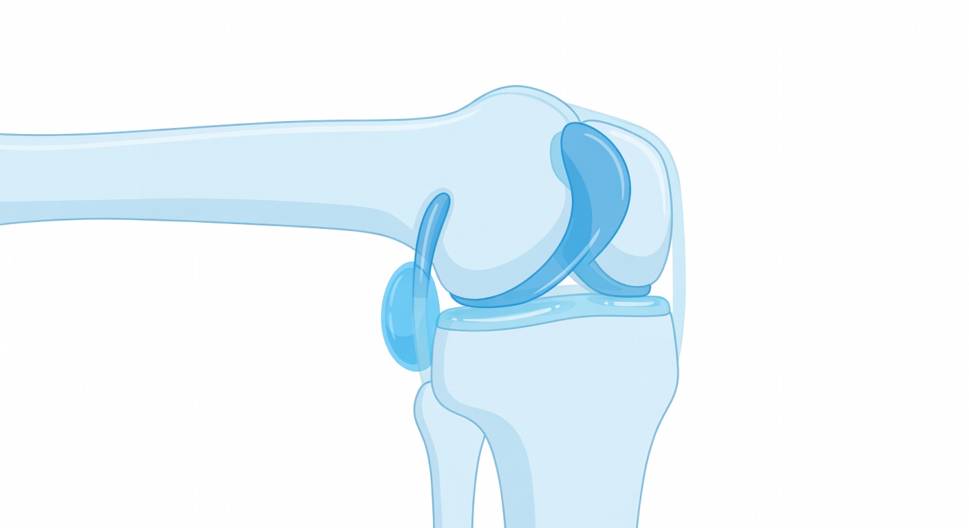
Each knee has two menisci: the medial meniscus on the inner side and the lateral meniscus on the outer side. These C-shaped pads of cartilage help spread body weight across the joint and improve stability.
A meniscus tear may happen suddenly with twisting, pivoting, or squatting. It can also develop gradually as the tissue becomes less flexible with age or mild wear-and-tear changes. MRI reports may describe a tear as horizontal, oblique, radial, complex, displaced, or nondisplaced. A nondisplaced tear means the torn piece has not clearly moved out of place.
Symptoms that can fit a meniscus tear
- Pain along the inner or outer joint line
- Swelling after activity or after an injury
- Catching, clicking, or a sense that something is moving inside the knee
- Difficulty squatting, twisting, or going up and down stairs
- True locking, where the knee gets stuck and cannot fully straighten
However, not every meniscus finding on MRI causes symptoms. Some tears are found in people whose pain is mainly from arthritis, tendon irritation, kneecap tracking problems, or another source. This is why clinicians compare the MRI with where the pain is, how the injury happened, and what movements reproduce symptoms.
Why reports may say “possible” meniscus tear
Radiologists look for abnormal signal that reaches the surface of the meniscus. Sometimes the finding is subtle, seen on only one image, or limited by motion, image quality, or missing sequences. In those cases, the report may say suspicious for or possible tear rather than definite tear.
A complete knee MRI usually includes images in several directions, commonly sagittal, coronal, and axial planes, with different sequences that highlight fluid, cartilage, bone marrow, and soft tissues. A single MRI series may show a clue, but it may not be enough to fully assess the menisci, ACL, PCL, collateral ligaments, and cartilage.
Small joint effusion: what does fluid mean?
A joint effusion means there is extra fluid inside the knee joint. A small amount of joint fluid can be a nonspecific finding. In plain language, it means the knee looks mildly irritated, but the MRI finding alone does not prove the cause.
Possible reasons for a knee effusion include a recent sprain or twist, meniscus irritation, cartilage wear, osteoarthritis flare, inflammatory arthritis, or less commonly infection or bleeding into the joint. The amount of fluid matters, but the story matters more. A small effusion after a long hike may mean something different than a rapidly enlarging swollen knee after a major injury.
Why “small effusion” is often not a diagnosis
- It does not identify one specific injury by itself.
- It may appear with both minor and more significant knee problems.
- It can be seen with arthritis or inflammation even without a new tear.
- It needs to be matched with pain location, swelling, warmth, injury history, and exam findings.
Baker cyst: fluid behind the knee
A Baker cyst, also called a popliteal cyst, is a pocket of joint fluid that collects at the back of the knee. It is often related to extra fluid inside the knee joint. The cyst itself is usually a sign of something irritating the knee rather than a separate disease.
Some Baker cysts cause no symptoms. Others may create fullness, tightness, or aching behind the knee, especially when bending or straightening the leg. If a cyst leaks or ruptures, it can cause calf discomfort and swelling that may mimic other conditions. Because symptoms in the calf can have different causes, new or concerning swelling should be medically evaluated.
Mild osteoarthritis on MRI
Osteoarthritis means wear-and-tear change in the joint. MRI reports may mention cartilage thinning, small bone spurs called osteophytes, or degenerative changes in one or more compartments of the knee. “Mild” osteoarthritis means these changes are present but not described as advanced.
Mild osteoarthritis may contribute to aching, stiffness, swelling after activity, or pain with stairs. It can also make meniscus tissue more likely to show degenerative tearing. In some people, MRI findings look more impressive than symptoms; in others, pain is significant even when imaging changes are described as mild.
Possible discoid lateral meniscus
A discoid lateral meniscus is a meniscus shape variant. Instead of the usual C shape, the lateral meniscus is broader and more disk-like. Some people have this their whole lives and never know it. In others, it may be associated with snapping, popping, catching, pain on the outer side of the knee, or a higher chance of tearing.
If an MRI report says “possible discoid lateral meniscus,” it usually means the shape looks broader than expected, but the full MRI and symptoms are needed to decide whether it matters.
Why symptoms and MRI do not always match
Knee pain is complex. A person can have a meniscus tear on MRI but pain from kneecap irritation. Another person can have a small tear that causes very specific catching or locking. Imaging is one part of the puzzle, not the whole answer.
Clinicians often ask: Did pain start after a twist? Is swelling immediate or delayed? Is pain on the inner or outer joint line? Is there true locking, or just stiffness? Is there instability or giving way? These details help decide whether an MRI finding is likely important.
Common next steps after these MRI findings
Treatment depends on the diagnosis, severity, activity goals, and exam. Many knee problems are first managed with nonsurgical care, such as activity changes, physical therapy, strengthening, anti-inflammatory strategies when appropriate, and time. Some situations, especially true mechanical locking, a displaced meniscus fragment, major ligament injury, or persistent symptoms despite conservative care, may lead to orthopedic or sports medicine evaluation.
The official radiology report matters. If only part of an MRI was reviewed, the final interpretation can change when all sequences are examined together.
When to talk to your doctor
Talk with a healthcare professional if knee pain, swelling, catching, locking, or instability is affecting your activities or not improving. Seek prompt care for severe pain, inability to bear weight, rapidly increasing swelling, fever, marked redness or warmth, new numbness or weakness, or calf swelling that is sudden or concerning.
This information is for general education and is not a personal medical diagnosis. Your doctor can connect your MRI findings with your symptoms and help decide what steps make sense for you.
Get AI-powered analysis of your CT or MRI scan
Upload your DICOM files and receive a clear, patient-friendly report in minutes.
Analyze my scan