Knee MRI: Effusion, Bone Bruise, ACL or Meniscus Tear?
Understand common knee MRI terms like effusion, bone bruise, ACL tear, and meniscus tear after injury or ongoing pain.

Have your own scan or report? Get a clear, plain-language explanation in minutes.
Why knee MRI reports can feel confusing
If you are limping, cannot fully bend your knee, or still have pain weeks after a fall or twist, an MRI report can raise as many questions as it answers. Words like joint effusion, bone contusion, suspected ACL injury, and possible meniscus tear may sound alarming, while phrases such as no displaced fracture or no macro tear may sound reassuring.
This article explains what these common MRI findings usually mean in plain language. It is general education, not a diagnosis, and it cannot replace a review of your full MRI, physical exam, symptoms, and medical history by a qualified clinician.
A knee MRI is not just one picture. It is a set of image sequences taken in different directions. Some injuries are obvious on one sequence, while others only become clear when all images are reviewed together.
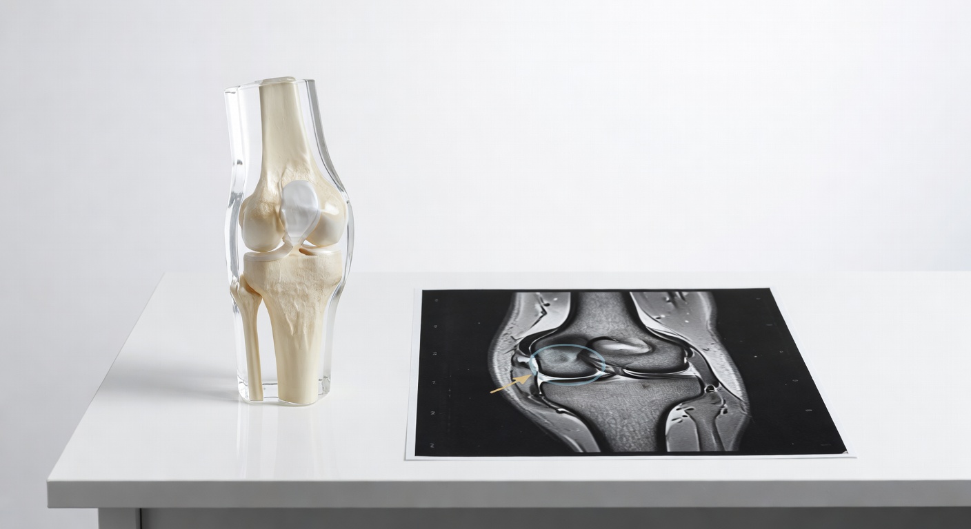
What does “joint effusion” mean?
A knee joint effusion means there is extra fluid inside the knee joint. Patients often describe this as swelling, tightness, pressure above the kneecap, or a feeling that the knee will not bend normally.
Fluid can build up for many reasons, including a recent injury, irritation of the joint lining, cartilage damage, arthritis, infection, or bleeding inside the joint. After a fall, twist, or sports injury, an effusion is a clue that something inside the knee has been irritated or injured, but it does not name the exact structure.
Small vs moderate-to-large effusion
MRI reports may describe the fluid as small, small-to-moderate, or moderate-to-large. A larger effusion can make the knee feel stiff and painful even if the report does not show a major tear. Fluid distends the joint capsule, and that pressure can limit bending and make walking uncomfortable.
That is why a person may say, “I cannot walk without limping” or “my bending is restricted,” even when the MRI impression sounds cautious rather than dramatic.
What is a bone bruise on MRI?
A bone bruise, also called an osseous contusion or marrow edema-like signal, means the inside of the bone shows swelling from impact or compression. It is not the same as a clearly displaced fracture, but it can still hurt significantly.
Bone bruises are often seen after twisting injuries, falls onto a straight leg, or contact injuries. They may appear in the outer part of the thighbone near the knee, called the lateral femoral condyle, or in the upper shinbone, called the tibial plateau.
Why lateral bone bruising can raise concern for ACL injury
When bone bruising is seen in the outer compartment of the knee, especially involving the lateral femoral condyle and the back or outer part of the tibial plateau, radiologists and sports medicine clinicians often look carefully at the anterior cruciate ligament, or ACL. This pattern can occur when the knee shifts or pivots during injury.
This does not automatically prove an ACL tear. It means the injury pattern makes an ACL sprain or tear more important to check. The full MRI, especially sagittal and coronal sequences, plus a hands-on stability exam, helps clarify whether the ACL is intact, partially torn, or completely torn.
Complete vs partial ACL tear: what does the MRI try to answer?
The ACL is one of the main stabilizing ligaments inside the knee. It helps control forward movement and rotation of the shinbone under the thighbone.
An MRI may describe the ACL as:
- Intact or continuous: the fibers can be followed from one attachment to the other.
- Sprain or increased signal: the ligament looks irritated or stretched, but fibers may still be present.
- Partial tear: some fibers appear disrupted, while others remain continuous.
- Complete tear: the ligament fibers are fully disrupted, not seen in normal position, or replaced by fluid and scar-like tissue.
Patients often ask, “Does the full MRI confirm an ACL tear?” or “Is it complete or partial?” Sometimes the report is clear. Other times, the wording is cautious, such as ACL injury cannot be excluded or suspicious for ACL sprain or tear. This usually means the available images or the specific sequence being reviewed do not fully settle the question.
If you have had previous ACL reconstruction, the report may use terms such as VKB Interponat or ACL graft. A phrase like continuous graft and no evidence of re-rupture is generally different from a new complete tear, but symptoms still need clinical correlation.
Does a reassuring report exclude a small meniscus tear?
Not always. The menisci are C-shaped cartilage cushions between the thighbone and shinbone. Meniscus tears can cause pain along the joint line, catching, swelling, difficulty squatting, or pain that “feels like a meniscus tear.”
MRI reports may say triangular configuration of the menisci, no macro tear, no displaced meniscal fragment, or no definite tear. These phrases are usually reassuring for a large or displaced tear. However, they may not always rule out subtle fraying, a tiny undersurface tear, or a tear that is difficult to see on one sequence.
Why all MRI sequences matter
A meniscus tear is usually assessed by looking for abnormal signal reaching the surface of the meniscus, changes in shape, missing tissue, displaced fragments, or associated cysts. This is easier when the radiologist can compare sagittal, coronal, and axial images with different MRI settings.
If only one series is available, such as a single coronal or sagittal sequence, the report may appropriately say that a tear is suspected or cannot be excluded. That is not the same as a full final answer.
What about cartilage and kneecap-stabilizing ligament injuries?
Cartilage covers the ends of bones and helps the joint glide smoothly. MRI may describe cartilage as normal, mildly worn, thinned, or having a chondral defect. A report saying there is no higher-grade chondral lesion is generally reassuring for major cartilage damage, but small cartilage changes can still be painful depending on location and the injury.
The kneecap, or patella, also has stabilizing structures, including the medial patellofemoral ligament on the inner side. If there has been a kneecap dislocation or subluxation, MRI may show bone bruises, cartilage injury, joint fluid, or soft-tissue swelling around the patella. If your symptoms include kneecap shifting, giving way, or pain around the front of the knee, your clinician may specifically review these structures.
Why do I still hurt if the MRI sounds “not too bad”?
Pain after knee injury can come from several sources, not just one dramatic tear. A moderate effusion can make bending difficult. A bone bruise can be painful with weight-bearing. Soft-tissue edema can reflect a sprain or contusion. A small meniscus or cartilage injury may be subtle. Muscle guarding after injury can also make the knee feel stiff, weak, or unstable.
Also, MRI findings and symptoms do not always match perfectly. Some people have severe pain with limited visible damage, while others have obvious MRI changes but fewer symptoms. This is why follow-up usually combines the report with your story: how the injury happened, whether the knee swelled quickly, whether it locks, whether it gives way, and whether walking or cycling increases pain.
Can I ignore the pain and return to sport or cycling?
It is understandable to ask whether you can keep cycling, train, or return to sport if the report does not show a major fracture. However, pain, swelling, limping, giving way, and restricted bending are signals that the knee is not back to normal function.
Return-to-activity decisions are not based on the MRI wording alone. They usually depend on your exam, range of motion, swelling, strength, stability testing, type of sport, and the final diagnosis. For example, a knee with suspected ACL injury or a painful effusion may need a different plan than a simple bruise or mild irritation.
Rather than “pushing through,” it is safer to have the MRI results interpreted in context by a clinician who can examine the knee and explain what activities are appropriate while healing continues.
What to expect at follow-up
At follow-up, your doctor, physiotherapist, or sports medicine clinician may:
- Compare the MRI report with the actual images, not just the impression.
- Check knee swelling, bending, straightening, and walking pattern.
- Test ACL, PCL, MCL, LCL, and meniscus signs.
- Ask whether the knee locks, catches, gives way, or feels unstable.
- Review whether prior surgery, such as ACL reconstruction, changes the interpretation.
- Discuss whether the MRI is definitive or whether specialist review is needed.
The goal of follow-up is not just to name the MRI finding, but to match the imaging with your symptoms and function.
When to talk to your doctor
Talk to a doctor or qualified clinician if you have ongoing pain after a fall or twist, cannot walk without limping, cannot fully bend or straighten the knee, have significant swelling, feel the knee giving way, or are unsure whether it is safe to return to sport. Seek urgent medical care for severe pain, fever, redness, numbness, major deformity, or inability to bear weight. This information is general education and is not a diagnosis or personal treatment advice.
Get AI-powered analysis of your CT or MRI scan
Upload your DICOM files and receive a clear, patient-friendly report in minutes.
Analyze my scan