Knee MRI: ACL Tear, Meniscus Tear, or Just Fluid?
Knee MRI reports can be confusing. Learn what fluid, ACL findings, meniscus tears, and bone bruises may mean.
Have your own scan or report? Get a clear, plain-language explanation in minutes.
Why knee MRI reports can feel confusing
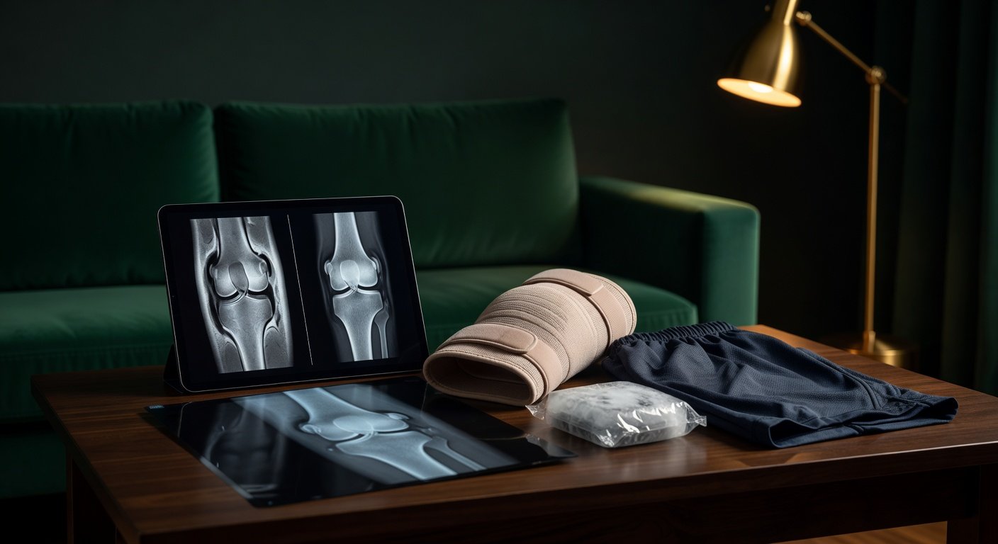
A knee MRI is often ordered when symptoms do not match a simple sprain: swelling, pain with walking, locking, catching, clicking, or a feeling that the knee may “give way.” But the report can still leave people unsure. One line may say there is a small joint effusion. Another may mention a possible meniscus tear, bone bruise, or abnormal ACL signal. Sometimes an AI review, clinic note, and official radiology report do not use the same words.
This article is general education, not a diagnosis. Knee MRI findings need to be matched with your injury story, physical exam, and the official radiologist report.
The key question is not only “What does the MRI show?” but also “Does this finding explain my symptoms?”
What does “fluid in the knee” mean?
A knee joint effusion means there is extra fluid inside the knee joint. A tiny amount of fluid can be normal. A small effusion can happen with irritation, inflammation, arthritis, or a mild injury. A moderate or large effusion, especially after a twist, fall, tackle, or pivot, raises more concern for an internal knee injury.
Fluid alone does not name the injury. It is a clue. It can appear with an ACL tear, meniscus tear, cartilage injury, bone bruise, inflammatory arthritis, or infection. The timing matters too. A knee that swells quickly after trauma may be viewed differently from a knee that slowly becomes puffy over weeks.
Fluid outside the joint is different. For example, swelling in front of the kneecap after a fall may be a bruised soft tissue collection, hematoma, seroma, or irritated prepatellar bursa. That is not the same as fluid deep inside the joint.
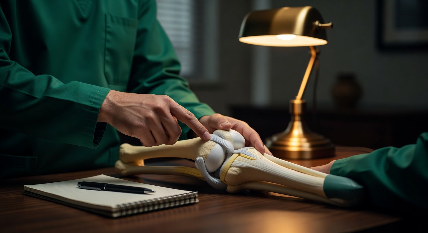
How an ACL tear can look on MRI
The anterior cruciate ligament, or ACL, helps control forward movement and rotation of the shin bone under the thigh bone. ACL injuries often happen with pivoting, cutting, landing, or contact injuries. People may describe a pop, swelling, instability, or giving way.
On MRI, a complete ACL tear may show loss of the normal tight, dark ligament fibers. The ACL may look wavy, swollen, poorly defined, or interrupted. Sometimes the MRI also shows a typical pattern of bone bruising in the outer part of the knee, often related to a pivot-shift type injury.
However, not every ACL injury is obvious. Some reports may say sprain, partial tear, high-grade tear, or complete rupture. After ACL reconstruction, the graft can be harder to judge because of surgical tunnels, metal artifact, scar tissue, or graft remodeling. In those cases, the physical exam is especially important.
Why the Lachman and pivot-shift tests matter
A clinician can test knee stability with maneuvers such as the Lachman test, anterior drawer test, and pivot-shift test. These tests ask a different question than MRI. MRI asks, “What do the tissues look like?” The exam asks, “How stable is the knee when it moves?”
A knee can have an ACL that looks abnormal but functions better than expected, or an MRI that is uncertain while the exam strongly suggests instability. Treatment decisions should not be based on one MRI phrase alone.
Meniscus tears: why subtle tears can be missed or debated
The menisci are C-shaped cartilage pads that help cushion and stabilize the knee. A meniscus tear may cause joint-line pain, swelling, catching, clicking, or pain with twisting. A displaced tear can sometimes cause true locking, where the knee cannot fully straighten or bend.
Radiologists look for meniscus signal that reaches the joint surface, abnormal shape, displaced fragments, or tears such as horizontal, oblique, radial, root, or bucket-handle tears. Some tears are very small. A small radial tear along the inner free edge of the meniscus can be difficult to see, especially if it appears on only one slice or one imaging plane.
This is one reason reports can differ. One reader may call a finding a subtle tear. Another may call it degenerative signal, fraying, or no definite macro-tear. MRI is powerful, but it is still interpreted by matching multiple image sequences and planes.
Do locking and catching prove a meniscus tear?
No. Locking, catching, clicking, and popping can come from several sources: a meniscus tear, cartilage flap, loose body, kneecap tracking issue, swelling, scar tissue, or pain-related muscle guarding. But persistent mechanical symptoms are important because they may change the urgency of orthopedic follow-up.
True locking usually means the knee physically cannot move through its normal range. Painful stiffness means movement is possible but limited by pain, swelling, or guarding. These feel similar to patients but may suggest different problems.
Bone bruises and why they matter
A bone bruise on MRI means there is injury-related swelling inside the bone marrow. It is not the same as a displaced fracture, but it can be painful and may point toward the mechanism of injury.
For example, a bone bruise in the lateral femoral condyle and posterolateral tibia can be seen with pivot-type injuries and may raise suspicion for an ACL injury. A bone bruise near the tibial tubercle in a growing athlete may fit traction irritation such as Osgood-Schlatter-type change, depending on the pain location and age.
Bone bruises can also make walking painful even when ligaments and menisci are not clearly torn. That is why “no fracture” does not always mean “nothing painful is present.”
When the official report differs from an AI review
AI tools may summarize MRI series quickly, but they are not a final diagnosis. They may overcall uncertain findings, miss subtle tears, or struggle when only some sequences show an abnormality. Official radiology reports are written after a radiologist reviews the full exam in context.
If an AI review says “possible meniscus tear” but the report says “no macro-tear,” that does not always mean one is wrong. It may mean the finding is too subtle or uncertain to call definite. If the clinic later identifies a small tear, it may have been visible only in a targeted review after symptoms and exam findings were known.
Helpful questions to ask include:
- Was the ACL continuous and taut, or was there concern for partial or complete tear?
- Was there a meniscus tear, fraying, or only nonspecific signal?
- Is the fluid inside the joint, in a bursa, or in the soft tissues?
- Are there bone bruises that suggest a pivot injury?
- Do my exam findings match the MRI report?
Why symptoms still matter after a “reassuring” MRI
A mostly normal MRI can be reassuring, but it does not automatically mean the knee is ready for sports. Pain with normal walking, worsening symptoms after a fall, giving way, limited bending, or inability to straighten the knee should be taken seriously. Sometimes swelling and pain come from bone bruising, bursitis, tendon irritation, cartilage injury, scar tissue, or a subtle meniscus injury that is difficult to see.
Likewise, an abnormal MRI does not automatically mean surgery is required. Many knee problems are first treated with activity modification, physical therapy, bracing, anti-inflammatory strategies when appropriate, and time. Decisions about surgery depend on the exact injury, age and activity goals, instability, mechanical symptoms, and exam findings.
When to talk to your doctor
Talk with your doctor, orthopedist, sports medicine clinician, or physical therapist if your MRI mentions an ACL injury, possible meniscus tear, bone bruise, or joint effusion and you still have pain, swelling, locking, catching, giving way, or trouble walking.
Seek prompt medical care if you have a hot red knee, fever, rapidly increasing swelling, severe worsening pain, inability to bear weight, numbness, loss of circulation, or a knee that is truly locked and cannot move. This information is for general education only and cannot diagnose your specific knee injury.
Get AI-powered analysis of your CT or MRI scan
Upload your DICOM files and receive a clear, patient-friendly report in minutes.
Analyze my scan