Kidney Cysts on CT or MRI: When Are They Benign?
Kidney cysts are common on CT and MRI. Learn what makes a cyst look benign and when more imaging may be suggested.
Have your own scan or report? Get a clear, plain-language explanation in minutes.
Why kidney cysts show up so often on scans
Kidney cysts are fluid-filled sacs in or near the kidney. They are a common incidental finding, meaning they are often seen on a CT or MRI done for another reason, such as abdominal pain, cancer staging, urinary symptoms, or a check of blood vessels.
Many kidney cysts are harmless. A report may describe them as simple cysts, parapelvic cysts, cortical cysts, or incompletely characterized cystic lesions. Those phrases can sound alarming, but they often reflect how much information the radiologist had from the scan, not necessarily that something dangerous was found.
This article is general education and is not a diagnosis. Your own scan should be interpreted by a radiologist who can review all available images, prior studies, symptoms, and lab results.
What makes a kidney cyst look benign?
Radiologists look for specific features that help separate a simple, benign cyst from a more complex cyst or a solid kidney mass. The most reassuring findings are:
- Fluid-like appearance: On CT, a simple cyst looks very low density, like water. On MRI, it usually follows fluid signal, often bright on T2 images.
- Thin, smooth wall: A simple cyst has a very thin wall and does not look thick, irregular, or lumpy.
- No solid part: There should be no internal nodule or solid tissue growing inside the cyst.
- No suspicious enhancement: After contrast, a simple cyst should not take up contrast in its wall or inside. Enhancement means tissue is gaining contrast, which can be more concerning.
- No complex internal features: Simple cysts usually do not have thick septations, heavy irregular calcification, blood products, or debris.
Size alone does not decide whether a cyst is benign. A small cyst can be simple, and a large cyst can also be simple. Radiologists care more about the cyst’s structure and whether it enhances after contrast.
Simple, parapelvic, and cortical cysts: what the words mean
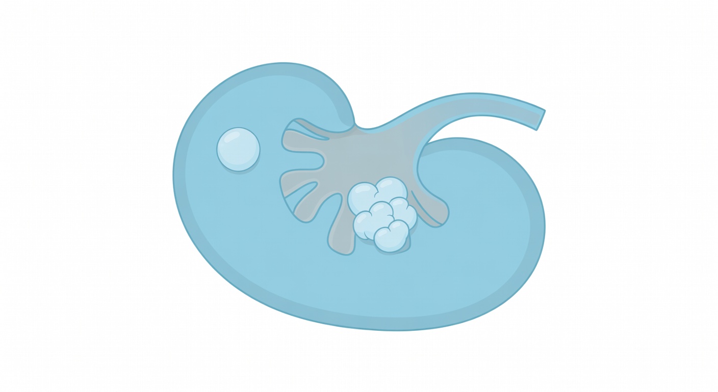
Simple kidney cyst
A simple kidney cyst is a smooth, fluid-filled cyst without suspicious features. If a report clearly calls a cyst simple, it usually means the imaging appearance is reassuring.
Cortical cyst
The kidney cortex is the outer part of the kidney. A cortical cyst is simply a cyst located in that outer kidney tissue. Many simple cysts are cortical cysts.
Parapelvic cyst
A parapelvic cyst is located near the center of the kidney, close to the urine-collecting system. These cysts can sometimes look like swelling of the kidney collecting system, called hydronephrosis. This is why ultrasound, delayed CT images, or comparison with other MRI sequences may be used to confirm whether the finding is a cyst or true urine blockage.
Why a report may say incompletely characterized
Sometimes a CT or MRI report says a kidney cyst is incompletely characterized or not fully evaluated. This does not automatically mean cancer. It often means the scan was not designed specifically to study the kidneys.
For example, a radiologist may only have one CT phase, such as an arterial or venous phase, instead of a full kidney protocol with images before and after contrast. Or an MRI may include only one sequence, without the T2, diffusion, pre-contrast, and post-contrast series needed to fully describe a cyst.
When only one series is available, the radiologist may be able to say the finding is probably a cyst but may avoid calling it definitely simple. That careful wording is meant to be accurate, not to cause panic.
Why ultrasound or comparison with old scans may be mentioned
Ultrasound is often useful because it can show whether a finding is filled with clear fluid and whether there is blood flow in a solid component. It is also widely available and does not use radiation. For a cyst that looks likely benign but is not fully characterized on CT or MRI, ultrasound may help confirm simple features.
Prior scans are also helpful. If a cyst looked the same months or years earlier, that stability is reassuring. If a cyst has changed, developed thick walls, new solid tissue, or new enhancement, the radiologist may recommend closer evaluation.
When kidney cysts need more attention
Some cysts are called complex because they have features beyond a simple fluid sac. Depending on the appearance, the report may recommend a kidney ultrasound, a dedicated renal CT or MRI protocol, follow-up imaging, or urology review.
Features that may lead to more evaluation include:
- Thick or irregular walls
- Internal septations, especially if thick or enhancing
- A solid nodule or mass-like component
- Enhancement after contrast
- Unclear difference between a parapelvic cyst and urine blockage
- Associated hydronephrosis, stones, or a concerning change from prior imaging
Radiologists may use a structured system, often called the Bosniak classification, to describe cystic kidney masses on CT or MRI. The exact category depends on detailed imaging features, especially enhancement.
Can kidney cysts cause flank pain?
Most simple kidney cysts do not cause pain. When people have flank or back pain and a cyst is found, the cyst may be unrelated. However, a cyst can sometimes contribute to symptoms if it is very large, stretches the kidney capsule, bleeds, ruptures, becomes infected, or blocks urine drainage. Those situations are less typical than an uncomplicated simple cyst.
Can cysts cause blood in the urine?
A simple cyst usually does not cause blood in the urine. Blood in the urine, also called hematuria, can come from many causes, including stones, infection, prostate conditions, kidney disease, bladder problems, or tumors. If hematuria is present, doctors usually evaluate it based on the full clinical picture rather than assuming a simple cyst is the cause.
Can kidney cysts cause infections?
Most simple cysts are not related to urinary tract infections. Rarely, a cyst itself can become infected, which may cause fever, pain, and abnormal blood or urine tests. Recurrent urinary infections usually prompt doctors to look for other causes as well, such as stones, bladder emptying problems, obstruction, or other urinary tract conditions.
Do kidney cysts affect kidney function?
A few simple cysts usually do not affect kidney function. Kidney function is more likely to be affected by conditions such as chronic kidney disease, diabetes, high blood pressure, obstruction of urine flow, or inherited cystic kidney diseases. If a person has many cysts in both kidneys, enlarged kidneys, abnormal kidney blood tests, or a family history of kidney cystic disease, the doctor may consider a broader evaluation.
How to read the wording in your report
Here are common phrases and what they often mean:
- Simple cyst: Reassuring fluid-filled cyst features are seen.
- Likely cyst or probable cyst: The finding looks cystic, but the scan may not show every feature needed for certainty.
- Parapelvic cyst: A cyst near the kidney collecting system, sometimes needing distinction from urine blockage.
- No hydronephrosis: No clear swelling of the kidney from blocked urine flow.
- Incompletely characterized: More sequences, phases, ultrasound, or prior scans may be needed to describe it fully.
The most important step is to review the official report with the clinician who ordered the scan. They can connect the imaging words with symptoms, urine tests, kidney function blood tests, and medical history.
When to talk to your doctor
Talk to your doctor if your report mentions a complex cyst, enhancement, a solid component, hydronephrosis, or an incompletely characterized kidney lesion. You should also seek medical advice if you have blood in the urine, fever, repeated urinary infections, new or severe flank pain, trouble urinating, or abnormal kidney function tests. This information is for general education and cannot determine what your specific cyst means without a full medical review.
Get AI-powered analysis of your CT or MRI scan
Upload your DICOM files and receive a clear, patient-friendly report in minutes.
Analyze my scan