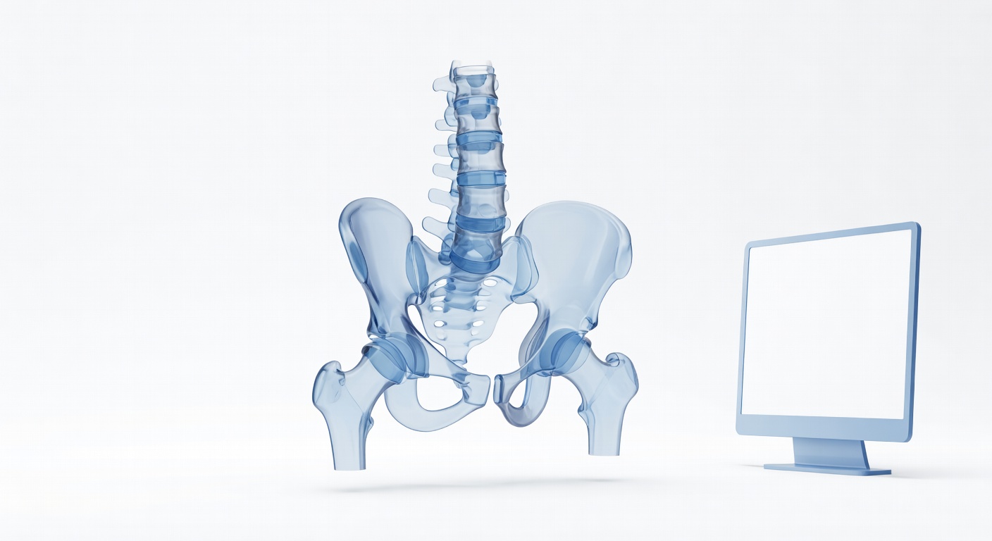
Degenerative Spine and Hip Changes on CT
CT and MRI often show spine or hip wear-and-tear changes. Learn what these terms mean and why they may not match pain.

Have your own scan or report? Get a clear, plain-language explanation in minutes.
This article is for general education only. It is not a diagnosis and does not replace the official radiology report or a visit with a qualified clinician.
Why spine and hip changes show up on CT scans
Many people have a CT scan of the abdomen, pelvis, kidneys, or hips for one reason and then notice unexpected words in the report: lumbar spondylosis, facet arthritis, pars defects, mild spondylolisthesis, hip osteoarthritis, or chronic degenerative changes. These findings can sound alarming, but they often describe long-term wear-and-tear changes rather than a new emergency.
CT scans are very good at showing bone detail. Even if the scan was ordered to look for abdominal pain, urinary stones, bleeding, or injury, the images often include the lower spine, pelvis, sacroiliac joints, and hips. Radiologists usually mention visible bone and joint changes because they are part of the scan, even when they are not the main reason for the test.
Incidental does not mean imaginary. It means the finding was seen while looking for something else, and it may or may not be related to your symptoms.
Common terms you may see in a report
Lumbar spondylosis
Lumbar spondylosis is a broad term for age-related or wear-related changes in the lower spine. It can include small bone spurs, disc space narrowing, thickened ligaments, and arthritis of the small joints in the back of the spine. It is similar to saying there are degenerative changes in the low back.
Facet arthritis or facet arthropathy
The facet joints are small joints at the back of each spinal level. They help guide movement and keep the spine stable. Over time, these joints can develop arthritis, just like knees, hips, or fingers. Facet arthritis can be associated with stiffness or aching low back pain, but it can also be present without symptoms.
Disc degeneration
Spinal discs sit between the bones of the spine and act like cushions. Reports may mention disc space narrowing, vacuum disc phenomenon, or degenerative disc disease. Despite the word disease, this often refers to long-term disc wear rather than an infection or cancer. CT can show disc height and bone changes, while MRI is better for discs, nerves, and soft tissues.
Pars defects and spondylolysis
A pars defect, also called spondylolysis, is a small break or gap in a specific part of a vertebra, most often at L5 near the base of the spine. It may come from stress over time and can be longstanding. Some people never know they have it. In others, it may contribute to low back pain, especially with extension-based activities.
Mild spondylolisthesis
Spondylolisthesis means one vertebra has slipped slightly forward or backward compared with the one below it. Reports often describe the grade, such as grade 1, meaning a mild slip. This can occur with pars defects or with arthritis-related weakening of the stabilizing joints and discs. Mild slips are often monitored in the context of symptoms, posture, nerve findings, and function.
Hip osteoarthritis
Hip osteoarthritis, sometimes called coxarthrosis, means the hip joint shows signs of cartilage wear and bone remodeling. CT or X-ray reports may mention joint space narrowing, bone spurs called osteophytes, hardening of bone under the cartilage called subchondral sclerosis, or small cyst-like changes. Hip osteoarthritis can cause groin pain, buttock pain, thigh pain, stiffness, or reduced walking tolerance, but imaging alone does not tell the whole story.
Why imaging severity does not always match pain severity
One of the most confusing parts of imaging is that the scan may look worse than the symptoms, or the symptoms may feel worse than the scan looks. Pain is influenced by many factors, including inflammation, nerve sensitivity, muscle strength, sleep, stress, previous injuries, and how the body is being used day to day.
For example, a CT may show moderate hip osteoarthritis in someone who walks comfortably. Another person may have severe hip or back pain with only mild changes on CT because the pain is coming from soft tissues, nerves, inflammation, or a problem that CT does not show well. MRI may be better when clinicians are concerned about nerve compression, bone marrow swelling, infection, certain fractures, or soft-tissue problems.
Imaging also captures a single moment in time. A report may describe chronic changes that developed slowly over years. Symptoms, however, can flare suddenly after a fall, a long drive, heavy lifting, illness, or a change in activity. That is why clinicians match the imaging with the story: where the pain is, what triggers it, what improves it, and whether there are nerve or strength changes.
How spine, hip, and leg symptoms can overlap
Low back, hip, buttock, groin, and leg symptoms can come from different structures that are close together. Hip joint arthritis often causes pain in the groin or front of the thigh, but it can also be felt in the buttock or knee. Lumbar spine problems may cause back pain, buttock pain, numbness, tingling, or pain traveling down the leg. Sacroiliac joint arthritis may cause pain near the back of the pelvis.
Because these patterns overlap, a CT finding does not automatically prove the source of pain. A physical exam, walking assessment, range-of-motion testing, and sometimes diagnostic injections or additional imaging may help a clinician decide which finding matters most.
General lifestyle themes that often help joint and spine health
There is no single plan that fits everyone, and treatment choices should be discussed with a clinician. In general, long-term spine and hip care often focuses on keeping the body as strong, mobile, and active as possible.
- Regular activity: Walking, cycling, swimming, or other low-impact movement can help maintain joint nutrition, circulation, and endurance.
- Strength training: Building the muscles around the hips, core, and legs may reduce stress on painful joints and improve stability.
- Mobility work: Gentle stretching and range-of-motion exercises may help with stiffness, especially when guided by a physical therapist.
- Weight management: For some people, reducing excess body weight can lower mechanical load on the hips, knees, and spine.
- Pacing: Alternating activity with rest and increasing exercise gradually may reduce flare-ups.
- Fall prevention: Good footwear, balance work, vision care, and home safety can be important, especially when CT reports mention bone thinning or chronic spinal changes.
Medications, injections, physical therapy, assistive devices, or surgery may be considered in selected situations, but decisions depend on symptoms, exam findings, overall health, and personal goals.
Reading your report with the right context
If your report says there is no acute fracture or no urgent abdominal problem but also lists degenerative spine or hip changes, both statements can be true. The scan may have ruled out the main emergency while also documenting chronic findings. It is reasonable to ask your clinician which findings are important, which are incidental, and whether any follow-up is needed.
Helpful questions include: Does this finding match where my pain is? Are there signs of nerve involvement? Would physical therapy be appropriate? Is MRI needed if symptoms persist or if nerve symptoms are present? Are the hip changes significant enough to consider orthopedic evaluation?
When to talk to your doctor
Talk with a clinician if back, hip, or leg pain is persistent, worsening, limiting walking, or not improving with general self-care. Seek prompt medical attention for new leg weakness, numbness in the groin or saddle area, loss of bladder or bowel control, fever with back pain, severe pain after a fall, inability to bear weight, unexplained weight loss, or rapidly worsening symptoms. Your doctor can interpret the CT or MRI findings in the context of your symptoms and decide whether further evaluation is needed.
Get AI-powered analysis of your CT or MRI scan
Upload your DICOM files and receive a clear, patient-friendly report in minutes.
Analyze my scan