
Contained Perforation From Diverticulitis: Is Surgery Next?
A contained perforation can often be treated without emergency surgery. Learn what CT wording means and what doctors check on follow-up imaging.
Have your own scan or report? Get a clear, plain-language explanation in minutes.
Why the word “contained” matters
Seeing “perforation” in a CT report can sound as though the bowel has burst and emergency surgery must be next. With diverticulitis, however, the details matter. A contained perforation is different from a large, freely leaking bowel perforation.
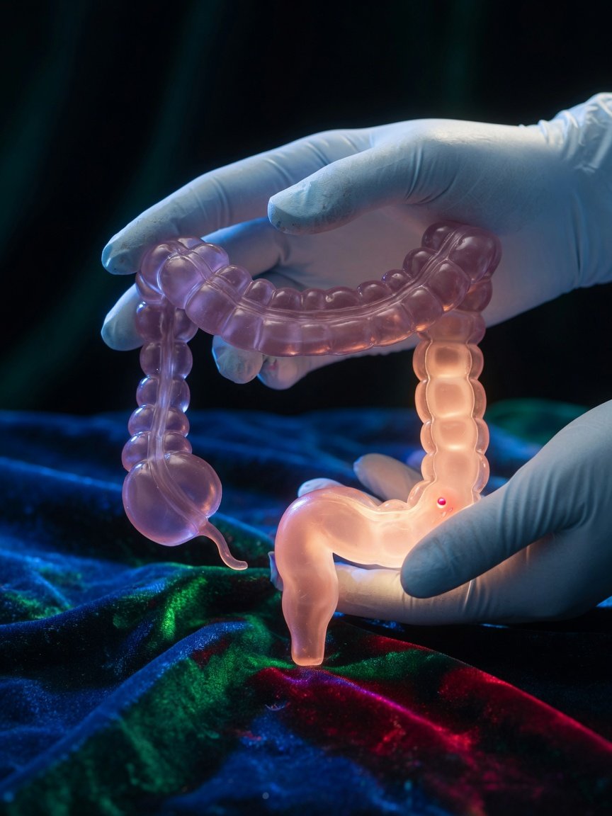
Diverticulitis occurs when small pouches in the colon, called diverticula, become inflamed. In some cases, a tiny opening develops in the wall of an inflamed pouch. A small amount of gas may escape, but nearby tissue and the body’s immune response can wall off the area. Radiologists may describe this as a microperforation, localized extraluminal gas, or contained perforation.
“Contained” generally means the leakage appears limited to a small area rather than spreading throughout the abdomen.
This distinction helps the medical team decide whether treatment can begin with close observation and medication or whether an urgent procedure is needed.
A typical CT-to-recheck timeline
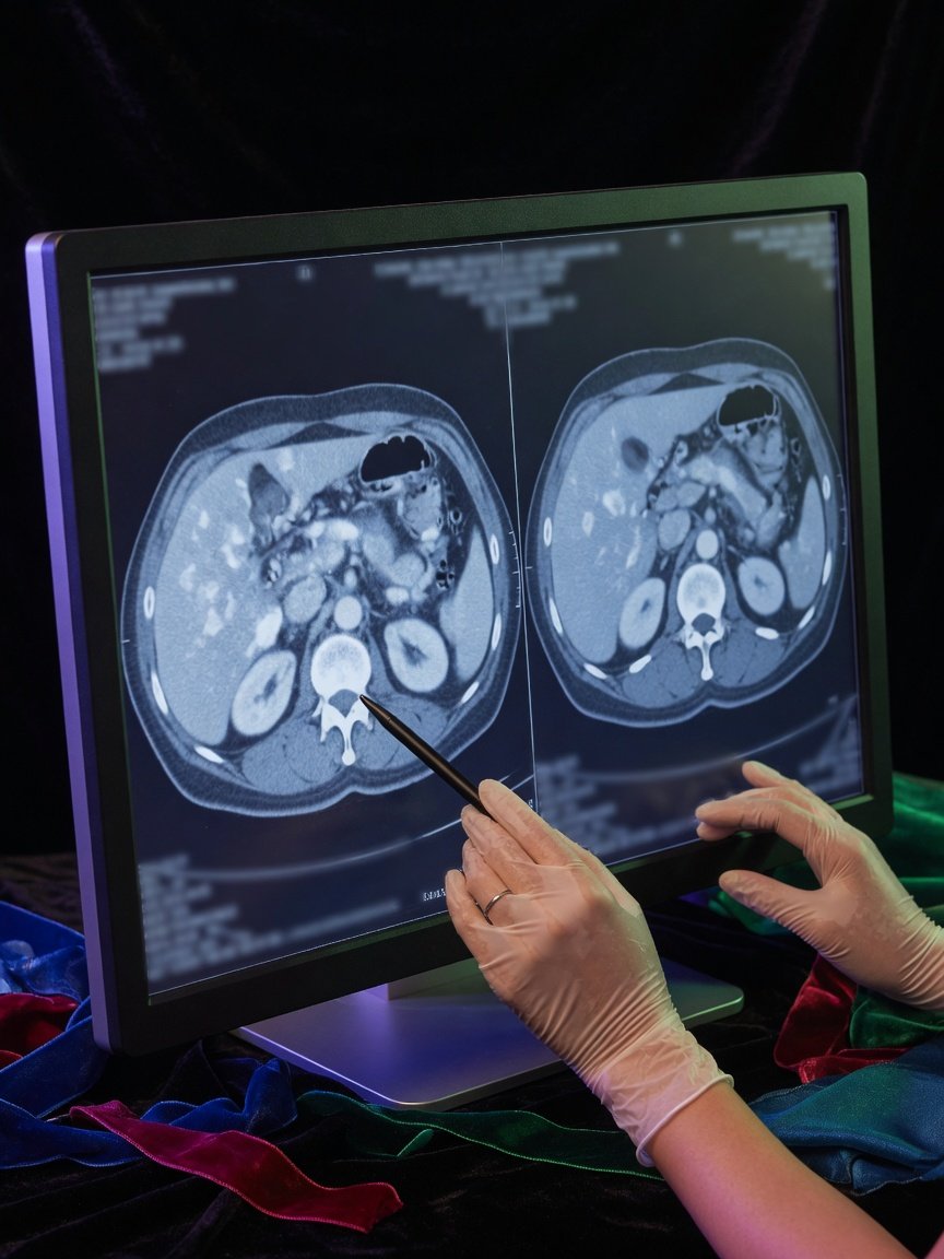
Consider the imaging pattern behind this article. An initial CT performed for abdominal and pelvic pain showed acute sigmoid diverticulitis. The sigmoid colon is the curved portion of the large intestine near the lower left side of the abdomen. The scan also showed a tiny nearby pocket of air interpreted as a contained perforation.
That finding can be serious enough to require hospital-level evaluation, but it does not automatically mean an operation. The immediate questions include:
- Is the person’s blood pressure and overall condition stable?
- Is the pain localized or spreading across the abdomen?
- Are there signs of sepsis or widespread abdominal infection?
- Is there a fluid-filled abscess that could require drainage?
- Can the person drink, take medication, and be monitored safely?
In this imaging sequence, another CT was obtained days later. It showed a small residual gas-containing area, while the surrounding inflammatory changes had resolved. The radiologist considered two possible explanations side by side: a residual collection related to the recent diverticulitis, or inflammation involving a separate small-bowel diverticulum. The report also identified which interpretation was favored.
That wording does not mean the other possibility is impossible. Radiologists often provide a differential diagnosis when findings can overlap. “Favored” means one explanation fits the location, appearance, and timeline better than the alternative. The treating clinician then compares that interpretation with symptoms, examination findings, laboratory results, and response to treatment.
Why antibiotics-first treatment is common
If a perforation appears small and contained, and the patient is clinically stable, treatment often starts without surgery. Depending on the severity and the person’s health, care may include:
- Antibiotics when clinicians believe bacterial infection or a complicated episode is present
- Fluids, sometimes given through a vein
- Pain and nausea treatment
- Temporary dietary changes based on tolerance
- Repeated abdominal examinations and monitoring
- Blood tests to follow inflammation and organ function
Some patients are monitored in the hospital, particularly when pain is severe, oral intake is difficult, or the CT suggests a complication. Others may be treated outside the hospital if their condition and support plan make that appropriate. The decision is individualized.
A contained leak may heal as inflammation settles and the body isolates the affected area. Avoiding emergency surgery can allow the colon to recover before any later decisions are made. It is important, however, to understand that “antibiotics first” does not mean “ignore it.” Close follow-up and attention to worsening symptoms remain essential.
What is the follow-up CT checking?
Not everyone with diverticulitis needs a short-interval repeat CT. Doctors are more likely to order one when symptoms persist, the first scan showed a complication, or the clinical course is not clear.
A recheck scan may look for:
- Less inflammation around the colon
- A shrinking or stable pocket of gas
- A developing fluid collection or abscess
- New or increasing free air
- Bowel blockage, a fistula, or another complication
- An alternative source of the original symptoms
A small amount of gas can remain visible even when surrounding inflammation has improved. Imaging does not always return to normal immediately. Doctors therefore interpret the scan as part of a trend rather than relying on one phrase in isolation.
Does a residual gas collection mean there is still a hole?
Not necessarily. Residual gas may remain in a walled-off space or within a nearby bowel diverticulum. Its significance depends on whether it is shrinking, whether fluid is present, and whether pain, fever, examination findings, and blood tests are improving.
Is every collection an abscess?
No. An abscess is usually an infected collection that often contains fluid and may also contain gas. A tiny gas pocket without a substantial fluid component may not be suitable for drainage. Location and change over time help distinguish an abscess from a bowel structure or a resolving contained leak.
When might a procedure or surgery be needed?
Surgery becomes more likely when there is widespread contamination of the abdomen, generalized peritonitis, unstable vital signs, uncontrolled infection, bowel obstruction, or failure to improve with nonoperative care. A larger accessible abscess may sometimes be treated with image-guided drainage rather than immediate colon surgery.
Even after recovery, an emergency operation is not automatically inevitable. A colorectal surgeon may discuss planned surgery after repeated attacks, persistent complications, narrowing of the colon, fistula formation, or a major effect on quality of life. The number of previous episodes is only one part of that decision.
Clinicians may also recommend colon evaluation after recovery, depending on the CT appearance, the person’s history, and when a high-quality colonoscopy was last completed. Colonoscopy is generally avoided during the most inflamed phase because the bowel is more vulnerable.
Reading the report without jumping to the worst conclusion
Pay attention to the complete impression rather than one alarming word. Terms such as tiny, localized, contained, improved, and resolved inflammation provide important context. So do less reassuring phrases such as increasing free air, drainable collection, or diffuse peritonitis.
A report explains what the images suggest; it does not determine treatment by itself. The most useful interpretation combines the scan with how the patient is doing in real time.
When to talk to your doctor
Ask your doctor what the collection represents, whether it is improving, and whether further imaging, colon evaluation, drainage, or a surgical consultation is expected. Seek urgent medical assessment for worsening or widespread abdominal pain, a rigid or very tender abdomen, persistent vomiting, fainting, confusion, fever with worsening illness, or inability to keep down fluids.
This article provides general education and cannot diagnose a condition or determine whether an individual needs surgery.
Get AI-powered analysis of your CT or MRI scan
Upload your DICOM files and receive a clear, patient-friendly report in minutes.
Analyze my scan