Cervical Spondylosis on MRI: Neck Arthritis Explained
Learn what common neck MRI terms mean, including disc bulges, bone spurs, stenosis, and mild cervical spondylosis.

Have your own scan or report? Get a clear, plain-language explanation in minutes.
What does cervical spondylosis mean?
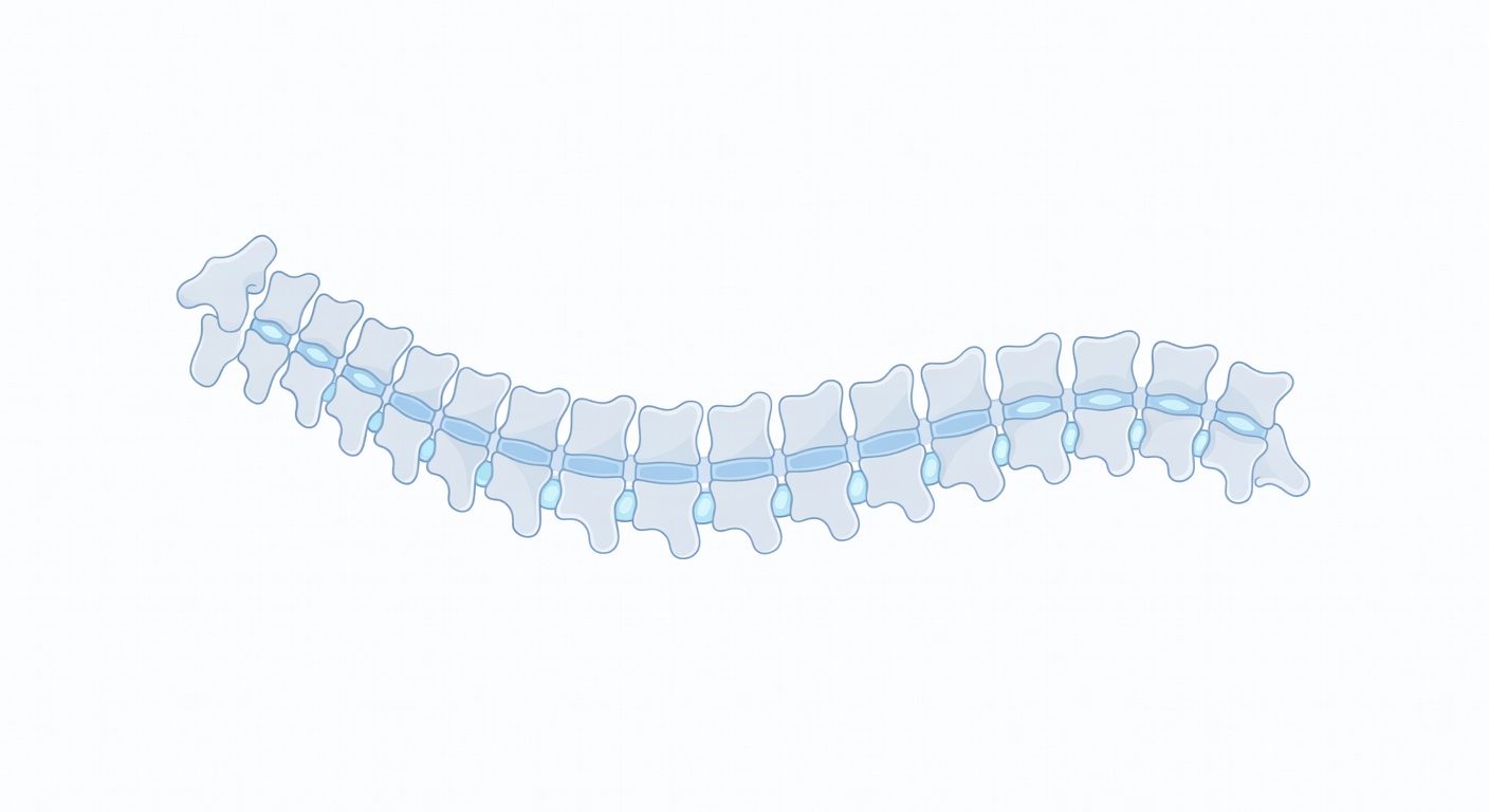
Cervical spondylosis is a medical term for age- and use-related changes in the neck part of the spine. The cervical spine includes the seven small bones in your neck, labeled C1 through C7. Between these bones are discs that act like cushions, and along the sides are joints and openings where nerves travel toward the shoulders, arms, and hands.
On MRI reports, cervical spondylosis may also be described as degenerative disc disease, disc osteophyte complex, uncovertebral or facet arthropathy, or simply wear-and-tear changes. These words can sound alarming, but mild to moderate degenerative findings are commonly seen on neck imaging, especially in the middle and lower neck levels such as C5-C6 and C6-C7.
This article is general education, not a diagnosis. Your own MRI findings need to be interpreted with your symptoms, physical exam, and the full official radiology report.
Why C5-C6 and C6-C7 are often mentioned
The lower neck does a lot of work. It helps support the head, allows bending and turning, and absorbs everyday stress from posture, movement, lifting, and screen use. Because of this, the discs and small joints around C5-C6 and C6-C7 are common places for degenerative changes to appear.
An MRI may show a disc that has lost some height or water content, a small bulge, or bone spurs along the edges of the vertebrae. These changes do not automatically mean there is a dangerous problem. The key question is whether the changes are pressing on a nerve root or the spinal cord, and whether that matches a person’s symptoms.
Common MRI terms in plain language
Disc bulge or disc protrusion
A disc bulge means the outer edge of a disc extends a little beyond its usual border. Think of a tire that is slightly flattened and spreading outward. A protrusion is a more focused outpouching. Many small bulges do not touch nerves or cause major narrowing.
Bone spurs and disc osteophyte complex
Osteophytes are bone spurs. They can form as the body responds to disc wear and joint stress. When a report says disc osteophyte complex, it usually means a combination of disc bulging and small bony overgrowths. These can gently indent the space around the spinal cord or narrow nerve openings.
Spinal canal stenosis
The spinal canal is the central tunnel that holds the spinal cord. Stenosis means narrowing. Mild or moderate canal stenosis means there is less room than usual, but it may still be enough space for the cord. Reports often note whether there is spinal cord compression or abnormal cord signal. If the report says there is no definite cord compression, that is generally a reassuring imaging detail, though symptoms still matter.
Foraminal narrowing
The neural foramina are side openings where nerve roots exit the spine. Foraminal narrowing means these openings are smaller than usual, often from disc changes, bone spurs, or arthritis in nearby joints. This is one MRI finding that may relate to arm symptoms, because a pinched nerve root can cause pain, tingling, numbness, or weakness along a nerve pathway.
Straightening of the neck curve
The neck normally has a gentle forward curve called cervical lordosis. MRI reports may mention straightening or loss of lordosis. This can be seen with muscle spasm, guarding from pain, positioning during the scan, or longer-term posture patterns. By itself, straightening is not a complete diagnosis.
Symptoms that may or may not come from neck arthritis
Cervical spondylosis can be present with no symptoms at all. When symptoms do occur, they can vary depending on which tissues are irritated.
- Neck pain or stiffness: often felt at the back of the neck or across the shoulders.
- Headache near the base of the skull: sometimes associated with neck muscle tension or joint irritation.
- Arm tingling or numbness: may occur if a nerve root is irritated or narrowed at the foramen.
- Arm pain: can travel from the neck into the shoulder, arm, or hand in a pattern that helps clinicians identify the involved nerve.
- Weakness or clumsiness: can be more concerning, especially if it is new, worsening, or affects walking or hand function.
It is important to remember that MRI findings and symptoms do not always match perfectly. A person can have mild-looking imaging findings with significant pain, or moderate-looking changes with few symptoms. That is why clinicians combine imaging with a neurologic exam, strength testing, reflexes, sensation checks, and a careful history.
What it means when there is no obvious spinal cord compression
Many MRI summaries say something like mild to moderate degenerative changes without high-grade spinal canal stenosis or cord compression. In plain language, this means the scan shows arthritis-like changes, but the spinal cord is not clearly being squeezed in a severe way on the available images.
This can be reassuring, but it does not rule out every source of pain. Nerve root irritation, muscle strain, joint inflammation, or posture-related stress may still contribute to symptoms. Also, some limited image sets may not fully show the nerve openings, so the official full MRI report is important.
Everyday prevention and self-care themes
No posture is perfect forever. The goal is not to hold your neck rigidly in one ideal position, but to reduce repeated strain and build comfortable movement habits.
- Change positions often: staying in one position for a long time can make the neck stiff, even if the posture looks good.
- Set screens near eye level: avoid prolonged looking down at a phone or laptop when possible.
- Support the arms: resting forearms on a desk or chair arms can reduce shoulder and neck tension.
- Use gentle movement breaks: slow neck turns, shoulder rolls, and walking breaks may help reduce stiffness for some people.
- Build strength gradually: physical therapy often focuses on deep neck muscles, shoulder blade control, and upper back strength.
- Sleep support matters: a pillow that keeps the neck comfortable and neutral may help some people wake with less stiffness.
These are general wellness ideas, not a personalized treatment plan. People with significant pain, neurologic symptoms, recent injury, or medical complexity should ask a clinician what activities are safe for them.
How doctors may approach treatment
Treatment depends on symptoms, exam findings, and the severity of nerve or cord involvement. Many people with mild to moderate cervical spondylosis start with non-surgical care. This may include education, activity changes, physical therapy, short-term pain-relief strategies, or targeted exercises. Some people may need additional testing or referral to a spine specialist, neurologist, orthopedic clinician, or neurosurgeon, especially when symptoms are progressive or neurologic deficits are present.
Surgery is not based on the word spondylosis alone. It is usually considered when there is significant nerve or spinal cord compression with matching symptoms, worsening neurologic findings, or pain that has not improved with appropriate conservative care. The right path varies from person to person.
When to talk to your doctor
Talk to your doctor if your MRI report mentions cervical spondylosis and you have persistent neck pain, arm pain, tingling, numbness, or stiffness that affects daily life. Seek prompt medical care for new or worsening arm or hand weakness, trouble walking, balance problems, loss of bladder or bowel control, severe pain after trauma, or rapidly spreading numbness. This article is for general education only and cannot diagnose your condition or replace care from a licensed clinician.
Get AI-powered analysis of your CT or MRI scan
Upload your DICOM files and receive a clear, patient-friendly report in minutes.
Analyze my scan