Bronchiectasis and Lung Nodules on CT: What They Mean
CT reports can sound alarming. Learn what bronchiectasis, mucus plugging, tree-in-bud changes, and small lung nodules often mean.
Have your own scan or report? Get a clear, plain-language explanation in minutes.
Why a chest CT report can feel confusing
A chest CT can describe many findings at once: bronchiectasis, mucus plugging, tree-in-bud opacities, emphysema, atelectasis, inflammatory consolidation, scarring, and small pulmonary nodules. These words can sound frightening, especially when the report recommends comparison with older scans or follow-up imaging.
This article is general education, not a diagnosis. CT findings must be interpreted by a radiologist and your clinician together with your symptoms, medical history, smoking history, infection history, and any prior imaging.
One of the most important questions after a CT is often not, “What is this word?” but “Is it new, changing, stable, or resolving?”
What is bronchiectasis?
Bronchiectasis means that some of the breathing tubes, called bronchi, are wider than usual. This widening is usually a chronic change. It can happen after repeated infections, inflammation, airway blockage, immune problems, aspiration from reflux or swallowing issues, or other lung conditions. Sometimes the exact cause is not clear.
On CT, bronchiectasis may be described as cylindrical, varicose, or cystic. These terms describe the shape and degree of airway widening. Bronchiectasis may be found in one area, such as a lower lobe, or in several areas, such as the right middle lobe, lingula, and lung bases.
People with bronchiectasis may have chronic cough, mucus or phlegm, repeated chest infections, wheezing, shortness of breath, fatigue, or coughing up blood. Others have mild CT findings with few symptoms. The CT appearance alone does not show how someone feels day to day.
Mucus plugging and bronchial wall thickening
Bronchiectasis can make it harder for the lungs to clear mucus. CT reports may mention mucus plugging or mucoid impaction, which means mucus is sitting inside small or medium-sized airways. Reports may also mention bronchial wall thickening, a sign that airway walls look irritated or inflamed.
These findings often fit with chronic airway disease. They can also be more noticeable during an infection or flare-up. If a person has fever, increasing cough, thicker or discolored sputum, worsening breathlessness, or low oxygen levels, clinicians may consider whether there is an active infection on top of chronic airway changes.
What does “tree-in-bud” mean?
Tree-in-bud opacities are tiny branching marks seen on lung CT. The name comes from their appearance: small airway branches can look like a budding tree. This pattern often points to inflammation or infection in the small airways, also called bronchiolitis.
Tree-in-bud changes can be seen with common respiratory infections, aspiration, chronic bronchiectasis-related inflammation, and sometimes less common or slower-growing infections. Some reports may mention atypical infection or nontuberculous mycobacteria as a possibility. That wording does not mean a diagnosis has been made. These infections usually require clinical assessment and, when appropriate, sputum testing or other microbiology tests.
Atelectasis, consolidation, inflammation, and scar: why reports use cautious language
CT reports may describe an area as atelectasis, consolidation, inflammatory opacity, or scarring. These can overlap in appearance.
- Atelectasis means part of the lung is partly collapsed or under-inflated. It can look like a band, streak, or patch.
- Consolidation means the air spaces look filled in, often from infection, inflammation, fluid, or other material.
- Inflammatory opacity is a broad term for a cloudy or denser area that may be related to irritation or infection.
- Scar or fibroatelectatic change suggests an older, more stable area from past inflammation, infection, or injury.
Radiologists often use cautious wording when an opacity could be infection, atelectasis, or scar, but a hidden underlying lesion cannot be fully excluded from one scan. This is why reports may recommend treatment correlation, comparison with old imaging, or a repeat CT to confirm that the area goes away or stays stable.
Emphysema and hyperinflation on CT
Some CT reports also mention emphysema, hyperinflation, or air trapping. Emphysema means there are areas where the tiny air sacs in the lungs have been damaged, often associated with smoking history but not limited to it. Hyperinflation means the lungs look over-expanded. Air trapping means some air remains in the lungs during breathing out, suggesting small airway disease.
These findings may be part of chronic obstructive pulmonary disease, commonly called COPD, but a diagnosis of COPD is not made from CT alone. Pulmonary function tests, symptoms, and clinical history are usually needed to understand how much the findings affect breathing.
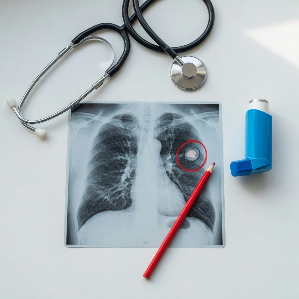
Small pulmonary nodules: what they usually mean
A pulmonary nodule is a small spot in the lung. Many nodules are found incidentally when CT scans are done for other reasons. A report may describe them as solid, subsolid, calcified, noncalcified, peripheral, or subcentimeter.
Small nodules can come from many causes, including old infection, inflammation, tiny scars, lymph nodes within the lung, or less commonly an early tumor. Calcified nodules or calcified lymph nodes often suggest old healed granulomatous infection, depending on the pattern and local medical context.
For lung nodules, management is usually based on:
- Size: exact measurement in millimeters matters.
- Appearance: solid, ground-glass, part-solid, smooth, spiculated, or calcified.
- Number: one nodule versus multiple small nodules.
- Location: upper-lobe nodules may be viewed differently from some lower-lobe nodules.
- Risk factors: smoking history, age, prior cancer, exposures, immune status, and family history may affect decisions.
- Prior imaging: a stable nodule over time is often managed differently from a new or growing nodule.
Many reports refer to guideline-based follow-up, such as local protocols or Fleischner-type recommendations. These guidelines help clinicians decide whether no follow-up, a repeat CT, or further testing is appropriate. The exact plan depends on the official radiology report and personal risk factors.
Why comparison with prior CT scans matters so much
When a CT report says “compare with prior imaging,” it is not just a formality. Older scans can show whether bronchiectasis has been long-standing, whether scarring is unchanged, whether an inflammatory opacity is new, or whether a small nodule has remained stable.
If a dense or irregular area resolves on follow-up, it is more likely to have been infection, inflammation, or atelectasis. If it persists or grows, clinicians may consider additional imaging, PET/CT, bronchoscopy, or biopsy depending on the full situation. This stepwise approach helps avoid both unnecessary alarm and missed important changes.
Helpful questions to ask after reading your CT report
- Are the bronchiectasis and airway changes mild, moderate, or severe?
- Do the findings fit my symptoms, such as cough, phlegm, fever, or shortness of breath?
- Is there mucus plugging or tree-in-bud change that suggests active infection or inflammation?
- Should sputum cultures or pulmonary function tests be considered?
- Is the opacity described as likely infection, atelectasis, scar, or something that needs follow-up?
- How large are the lung nodules, and were they measured on the original CT images?
- Are there old CT scans or chest X-rays available for comparison?
- If follow-up CT is recommended, what is the reason and what change are we looking for?
When to talk to your doctor
Talk to your doctor or the clinician who ordered the CT if your report mentions bronchiectasis, tree-in-bud opacities, mucus plugging, an irregular consolidation, or pulmonary nodules that need comparison or follow-up. A pulmonologist may be helpful when there is chronic cough, recurring infections, ongoing sputum, emphysema/COPD concerns, or uncertainty about infection versus scarring.
Seek urgent medical care for severe or worsening shortness of breath, chest pain, coughing up blood, persistent high fever, confusion, very low oxygen readings, or rapid worsening of symptoms. This article is for education only and cannot determine what your CT findings mean for you personally.

Get AI-powered analysis of your CT or MRI scan
Upload your DICOM files and receive a clear, patient-friendly report in minutes.
Analyze my scan