Breast MRI Enhancement and BI-RADS: What to Ask
Breast MRI enhancement, DWI bright spots, and BI-RADS scores can be confusing. Learn what they mean and what to ask next.
Have your own scan or report? Get a clear, plain-language explanation in minutes.
This article is for general education only. It is not a diagnosis and cannot replace review by your own doctor or a breast imaging radiologist.
Why breast MRI wording can feel alarming
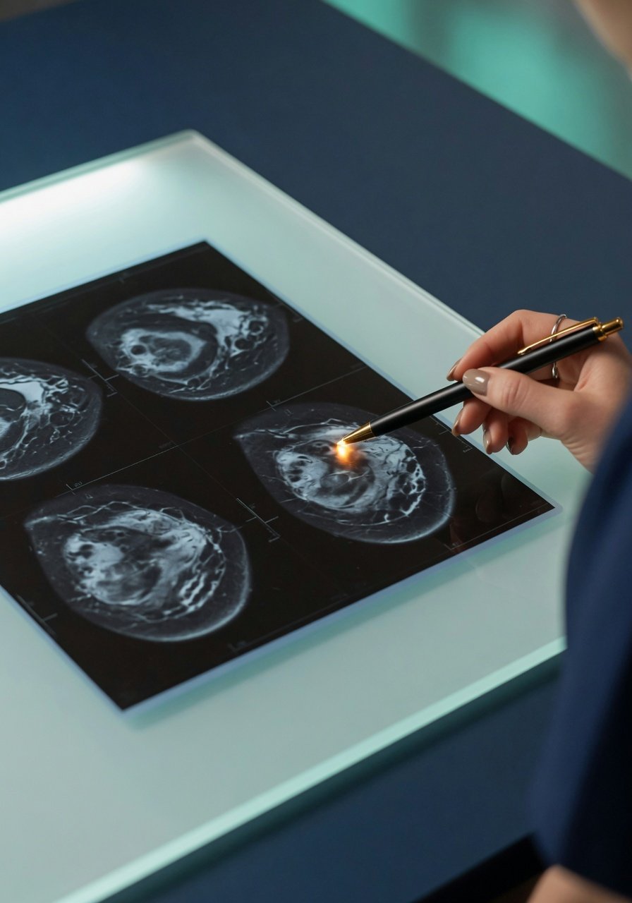
Breast MRI reports often use technical language: enhancement, DWI bright focus, ADC, non-mass enhancement, and BI-RADS 4B. It is understandable to wonder: Does this mean cancer? Is a bright spot dangerous? What if an AI summary sounds reassuring, but the formal report assigns a higher BI-RADS category?
The short answer is that no single word or sequence decides the meaning. Breast radiologists interpret the full MRI exam by looking across many image types, comparing with prior mammograms or ultrasounds, and matching the imaging with symptoms and physical exam findings.
A key question is not just, Is there enhancement? but What kind of enhancement is it, where is it, and does it match a real finding on other MRI sequences or ultrasound?
What does enhancement mean on breast MRI?
Breast MRI is commonly performed with contrast dye. Areas that take up contrast are described as enhancing. Enhancement happens because contrast travels through blood vessels and tissue spaces. Some enhancement is normal. Some is benign. Some patterns need closer evaluation.
Common types of enhancement
- Background parenchymal enhancement: Normal breast tissue may enhance, especially depending on hormones, menstrual cycle timing, medications, and breast density. If it is mild and symmetric, it is often less concerning.
- Mass enhancement: A discrete lump-like area enhances. Radiologists look at its shape, margins, internal pattern, and curve over time.
- Non-mass enhancement: Enhancement that does not form a round or oval mass. The pattern matters. Linear, segmental, clumped, or asymmetric patterns may be more concerning than scattered or symmetric background enhancement.
- Skin, nipple, chest wall, or lymph node enhancement: These areas are checked because involvement can change the level of concern and the next steps.
A report that says there is no definite suspicious enhancing mass or no suspicious non-mass enhancement is generally reassuring. But the final meaning still depends on the official BI-RADS assessment.
What about a DWI bright spot?
DWI stands for diffusion-weighted imaging. It is one part of a breast MRI that can show areas where water movement is more restricted. Some cancers can appear bright on DWI, but many non-cancer reasons can also cause brightness.
Radiologists do not look at DWI alone. They also check the ADC map. If a spot is bright on DWI but does not have a matching low ADC area, it may represent artifact, T2 shine-through, fluid, motion, or a non-specific finding rather than true restricted diffusion.
This is why a small DWI bright dot may be described as uncertain or not clearly suspicious if it has no matching abnormality on ADC and no matching suspicious enhancement. In that situation, a breast imaging specialist may review the original images slice by slice and decide whether it needs targeted ultrasound, short-term follow-up, or no special action beyond routine care.
BI-RADS: the category that guides next steps
BI-RADS is a standardized system used in breast imaging. It helps communicate how suspicious a finding is and what management is usually recommended. The category is usually more important than any single descriptive phrase.
- BI-RADS 0: Incomplete. More imaging or prior comparison is needed.
- BI-RADS 1: Negative. No concerning finding is seen.
- BI-RADS 2: Benign. A non-cancer finding is present.
- BI-RADS 3: Probably benign. Short-interval imaging follow-up is often recommended to confirm stability.
- BI-RADS 4: Suspicious. Biopsy is commonly considered. Subcategories 4A, 4B, and 4C reflect increasing concern.
- BI-RADS 5: Highly suggestive of malignancy. Tissue diagnosis is typically recommended.
- BI-RADS 6: Known biopsy-proven cancer.
If your report says BI-RADS 4B, that is not the same as a cancer diagnosis. It means the imaging finding is suspicious enough that the radiologist believes tissue sampling, such as a core needle biopsy, may be needed to know what it is. Benign results can still occur, including fibrocystic change, fibroadenoma, papilloma, inflammation, or post-procedure change, depending on the case.
Why MRI and ultrasound can disagree
It can be confusing when ultrasound shows one thing and MRI shows another. This does not always mean one test is wrong. They are different tools.
- MRI is very sensitive to contrast enhancement, blood flow patterns, and extent of disease. It can find areas that are hard to see on ultrasound.
- Ultrasound is excellent for evaluating a specific palpable lump, cystic versus solid features, ducts, and guiding biopsy.
- Mammography or tomosynthesis can show calcifications and architectural distortion that may not be obvious on ultrasound or MRI.
A small deep breast nodule, duct-related lesion, or post-surgical area may look different across tests. Prior history also matters. For example, someone with previous benign breast surgery or fibrocystic breast disease may have scars or benign changes that require careful comparison with older images.
When imaging results seem to conflict, the best next step is often not to choose one report over another, but to ask for radiology correlation: Does the MRI finding match the ultrasound finding? Is it in the same clock-face position and distance from the nipple? Has it changed over time? Can it be biopsied with ultrasound guidance, or is MRI-guided biopsy needed?
AI impressions versus the formal radiology report
Some patients now review AI-generated summaries before or alongside the official report. These summaries can help translate complex terms, but they are not the final diagnosis. An AI may summarize visible sequences as not showing a definite suspicious enhancement, while a human radiologist reviewing the full exam may identify a small irregular enhancing nodule and assign BI-RADS 4B.
In that situation, the formal radiologist report should guide medical decisions. If the difference is unclear, you can ask your clinician whether a breast imaging specialist can review the case again, compare all prior studies, or issue an addendum explaining the correlation.
Questions to ask your breast imaging specialist
Bringing specific questions can make the appointment more useful and less stressful.
About enhancement
- Does the full MRI show any enhancement of concern, or only mild background enhancement?
- Is the finding a mass, non-mass enhancement, or a skin/superficial lesion?
- What are the concerning features: irregular shape, unclear borders, spiculation, clumped pattern, or enhancement curve?
- Is there nipple, skin, chest wall, or lymph node involvement?
About DWI and ADC
- Does the DWI bright spot have a matching low ADC area?
- Could it be artifact, fluid, a cyst, or T2 shine-through?
- Does it match an enhancing lesion on the contrast images?
About BI-RADS and next steps
- What is the final BI-RADS category?
- If it is BI-RADS 3, when should follow-up imaging be done?
- If it is BI-RADS 4A, 4B, or 4C, what type of biopsy is recommended?
- Can the area be found on targeted ultrasound, or would MRI-guided biopsy be needed?
- How does this compare with my prior ultrasound, mammogram, MRI, or past biopsy results?
How to prepare for a follow-up visit
Before your appointment, gather prior imaging reports and, if possible, the actual images on disc or through an imaging portal. Write down any symptoms, such as a new lump, nipple discharge, skin dimpling, redness, focal pain, or recent procedures. Also note hormone therapy, menstrual cycle timing, pregnancy or breastfeeding status, and prior breast biopsies or surgeries.
It is reasonable to ask for plain-language clarification. For example: What exactly are we worried this could be, and what test will answer that question?
When to talk to your doctor
Talk with your doctor or breast imaging specialist if your report lists BI-RADS 0, 3, 4, or 5; if MRI and ultrasound results seem different; or if you have a new lump, nipple discharge, skin change, persistent focal pain, or a high-risk breast history. If a biopsy is recommended, your care team can explain why, which method is best, and how results will guide the next step.
Again, this information is general education and not a personal diagnosis. Your own care should be based on the official radiology report, prior imaging comparison, physical exam, and your clinician’s recommendations.
Get AI-powered analysis of your CT or MRI scan
Upload your DICOM files and receive a clear, patient-friendly report in minutes.
Analyze my scan