ACL Tears on MRI: Complete vs Partial Explained
Learn how MRI helps tell complete ACL tears from partial sprains, and why the full scan and knee exam matter.

Have your own scan or report? Get a clear, plain-language explanation in minutes.
Why ACL MRI reports can sound uncertain
After a pivot, tackle, twist, or giving-way injury, many people want a clear answer: is the ACL completely torn, partially torn, or intact? MRI is one of the best tools for looking at the anterior cruciate ligament, but the wording in reports can still feel frustrating. You may see phrases like suspicious for a high-grade tear, possible partial tear, or not definitive on this sequence alone.
This uncertainty often happens when only one image sequence is being discussed, or when the ACL looks abnormal but not enough information is visible to grade it confidently. The ACL runs diagonally through the center of the knee, so radiologists usually need multiple views and fluid-sensitive sequences to follow the fibers from top to bottom.
General education: This article explains common MRI language and injury patterns. It is not a diagnosis and cannot replace review of your full MRI by a radiologist or an exam by a clinician.
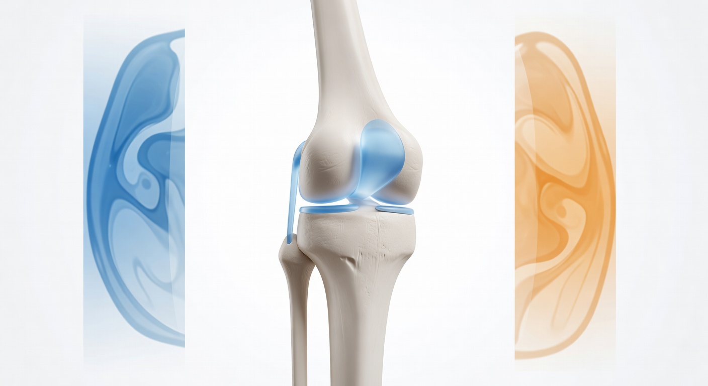
What the ACL normally looks like on MRI
The ACL is a strong band of tissue that helps stop the shin bone from sliding forward and helps control rotation of the knee. On MRI, a healthy ACL usually appears as a dark, tight, rope-like structure in the intercondylar notch, the space between the rounded ends of the thigh bone.
Radiologists look for whether the ligament is continuous, tight, and in the expected position. They also compare the ACL with nearby structures, especially the posterior cruciate ligament, menisci, cartilage, bone marrow, and joint fluid.
Complete ACL tear vs partial ACL tear on MRI
Signs that suggest a complete ACL tear
A complete ACL tear means the ligament fibers are fully disrupted enough that the ACL no longer acts as a continuous stabilizing band. MRI signs may include:
- Fiber discontinuity: the dark ACL band is interrupted or cannot be followed normally.
- Poor visualization: the ACL is hard to see because torn fibers are replaced by fluid and swelling.
- Abnormal high signal: fluid-sensitive images show bright signal within the ligament area, suggesting injury.
- Loss of normal taut shape: instead of a tight band, the fibers may look wavy, lax, frayed, or absent.
- Abnormal orientation: the ligament may not follow its usual diagonal course.
Reports may describe this as a high-grade tear, complete tear, rupture, or ACL not well visualized with fiber discontinuity. Sometimes a radiologist says “high-grade to complete” when the ACL is severely injured but a few fibers or scar-like tissue make exact grading difficult.
Signs that suggest a partial ACL tear or sprain
A partial tear means some ACL fibers are injured, but some fibers still appear continuous. A sprain may mean the ligament is stretched or swollen without a definite full-thickness break. MRI signs may include:
- Thickening or swelling of the ACL.
- Increased signal inside the ligament, meaning it looks brighter than usual on fluid-sensitive images.
- Some intact fibers that can still be traced from the femur to the tibia.
- Only part of the ligament bundle involved, while another portion remains dark and tight.
Partial ACL injuries can be difficult to grade because the ACL is made of fiber bundles that twist along an oblique path. One part may look injured on one image, while another view shows remaining intact fibers.
Why one MRI sequence may be suspicious but not definitive
MRI scans are made of several sequences, each designed to highlight different tissues. For ACL injuries, radiologists commonly use sagittal, coronal, and axial images, often with proton density or T2 fat-suppressed sequences. Some MRI protocols also include an ACL-focused oblique sequence.
A single sequence can raise concern but may not prove the grade of tear. For example:
- Sagittal images often show the ACL well, but may miss details if the ligament is not perfectly aligned with the slice.
- Coronal images can show central notch signal and side ligament injuries, but may not show the full ACL length.
- Axial images can show swelling, joint fluid, and bone bruise patterns, but are usually not enough by themselves to grade the ACL.
- Highly processed or 3D reformatted images may help in some settings but may not replace standard diagnostic MRI sequences.
This is why a report may say the ACL is indistinct or suspicious for tear on one sequence, while recommending correlation with the complete MRI examination. In plain language, that means: the picture raises a real concern, but the full set of images is needed before calling it partial, complete, or intact.
How swelling, joint fluid, and bone bruising fit the pattern
Many ACL injuries come with extra fluid inside the knee, called a joint effusion. After a sudden twisting injury, the knee may swell because of bleeding or inflammation inside the joint. MRI reports may describe a small, moderate, or large effusion, often in the suprapatellar recess above the kneecap.
Bone bruising is another important clue. With pivot-shift type injuries, MRI may show bright marrow signal in the lateral femoral condyle and the posterolateral tibial plateau. This pattern can happen when the bones briefly impact each other during the twist. Bone bruising does not automatically prove a complete ACL tear, but it supports the idea of a significant internal knee injury.
Other associated findings may include meniscus tears, cartilage injury, low-grade MCL sprain, or a small popliteal cyst. Locking, catching, clicking, and giving-way symptoms can come from several structures, so the full MRI review matters.
Does MRI “confirm” whether the ACL is intact?
Sometimes it can. If the ACL is clearly continuous, dark, tight, and normally oriented on multiple sequences, the report may say it is intact. If the fibers are clearly disrupted and the ligament cannot be followed, the report may diagnose a complete tear.
But there is a middle zone. A ligament can be swollen and abnormal without a clean visible gap. Scar tissue, motion blur, slice angle, fluid, and partial-volume effects can make the ACL look unclear. In those cases, the report may use cautious language such as suspected ACL sprain or partial tear or suspicious for high-grade tear.
Why the physical exam matters
MRI shows structure, but the knee exam shows function. Orthopedic clinicians often use tests such as the Lachman test, anterior drawer test, and pivot shift test to assess ACL stability. A person with a partial tear may still have instability, while another person with abnormal MRI signal may have a stable-feeling knee.
This is why grading an ACL injury is not based on one image alone. The most useful answer comes from combining:
- the full MRI protocol, not just one picture sequence;
- the official radiology report;
- the mechanism of injury, such as pivoting or a tackle;
- symptoms like swelling, locking, catching, clicking, and giving way;
- hands-on stability testing by a qualified clinician.
For Spanish-speaking patients asking, “¿puedes con todas las pruebas graduar el desgarro?” the key idea is: grading usually requires all MRI sequences plus the physical exam. Imaging can strongly suggest the grade, but the final clinical interpretation depends on the whole picture.
When to talk to your doctor
Talk with a doctor, sports medicine clinician, or orthopedic specialist if you have ongoing giving-way, significant swelling, trouble bearing weight, locking, or a report that says possible, high-grade, or complete ACL tear. Ask them to review the full MRI, not just one sequence, and to explain how the imaging matches your knee stability exam.
Get AI-powered analysis of your CT or MRI scan
Upload your DICOM files and receive a clear, patient-friendly report in minutes.
Analyze my scan