
SI Joint MRI: Sacroiliitis, Wear, or Osteitis Condensans?
Mild SI joint changes do not always mean rheumatic disease. Learn what MRI findings can—and cannot—explain about back, buttock, and leg pain.
Have your own scan or report? Get a clear, plain-language explanation in minutes.
Does an abnormal SI joint MRI mean inflammatory arthritis?
Reading about “sclerosis,” “chronic changes,” or “bone marrow edema” on an MRI can be unsettling, especially when pain travels from your sacrum or buttock into your groin or leg. Do these findings mean a rheumatic disease? Are they the reason you hurt?
Not necessarily. Several conditions can produce similar-looking changes near the sacroiliac, or SI, joints. The pattern of findings matters more than any single word. Mild changes may contribute to pain, be unrelated to it, or explain only part of the symptoms.
This article provides general education, not a diagnosis or individual medical advice. Imaging needs to be interpreted alongside symptoms, examination findings, and the official radiology report.
What the SI joints are—and what MRI looks for
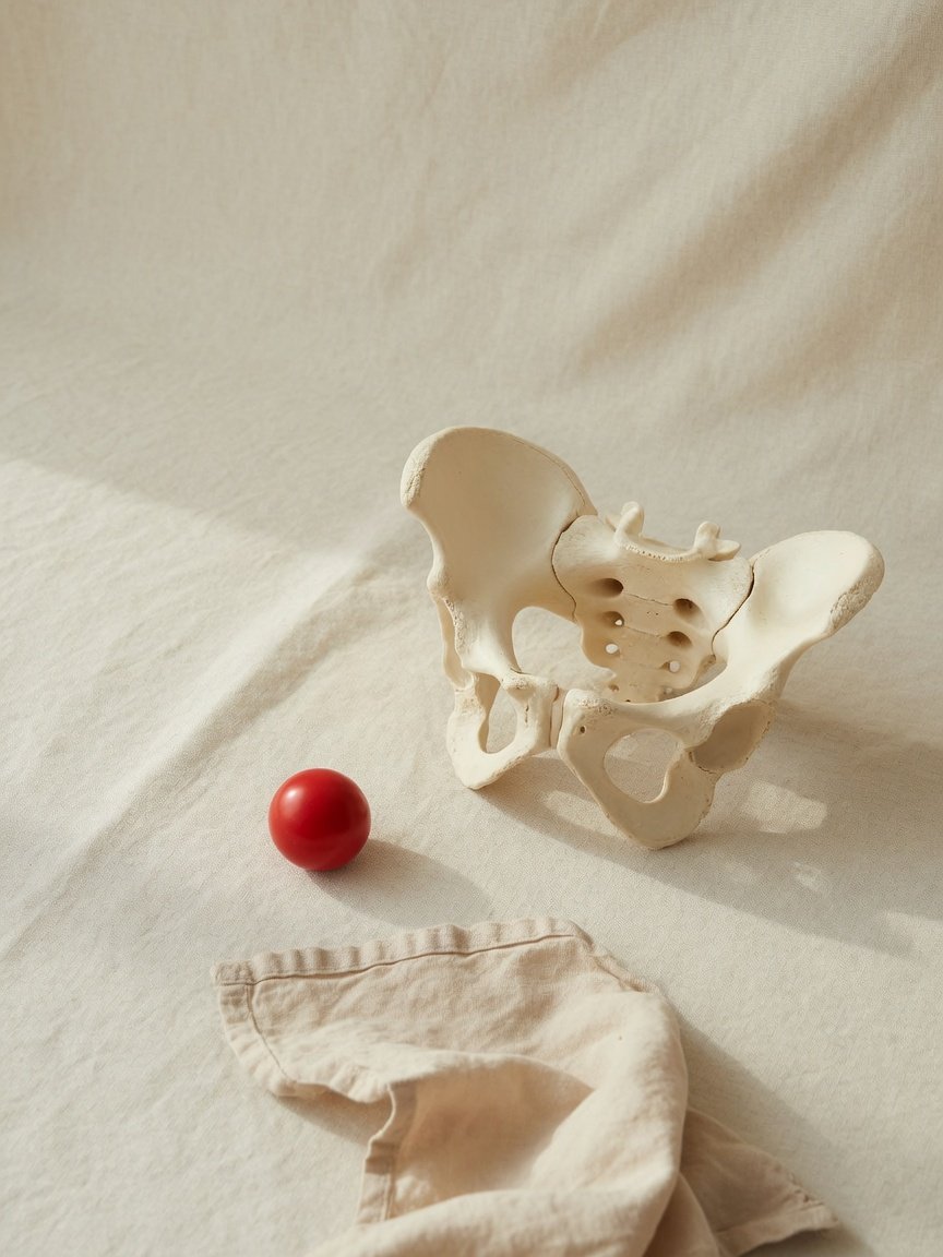
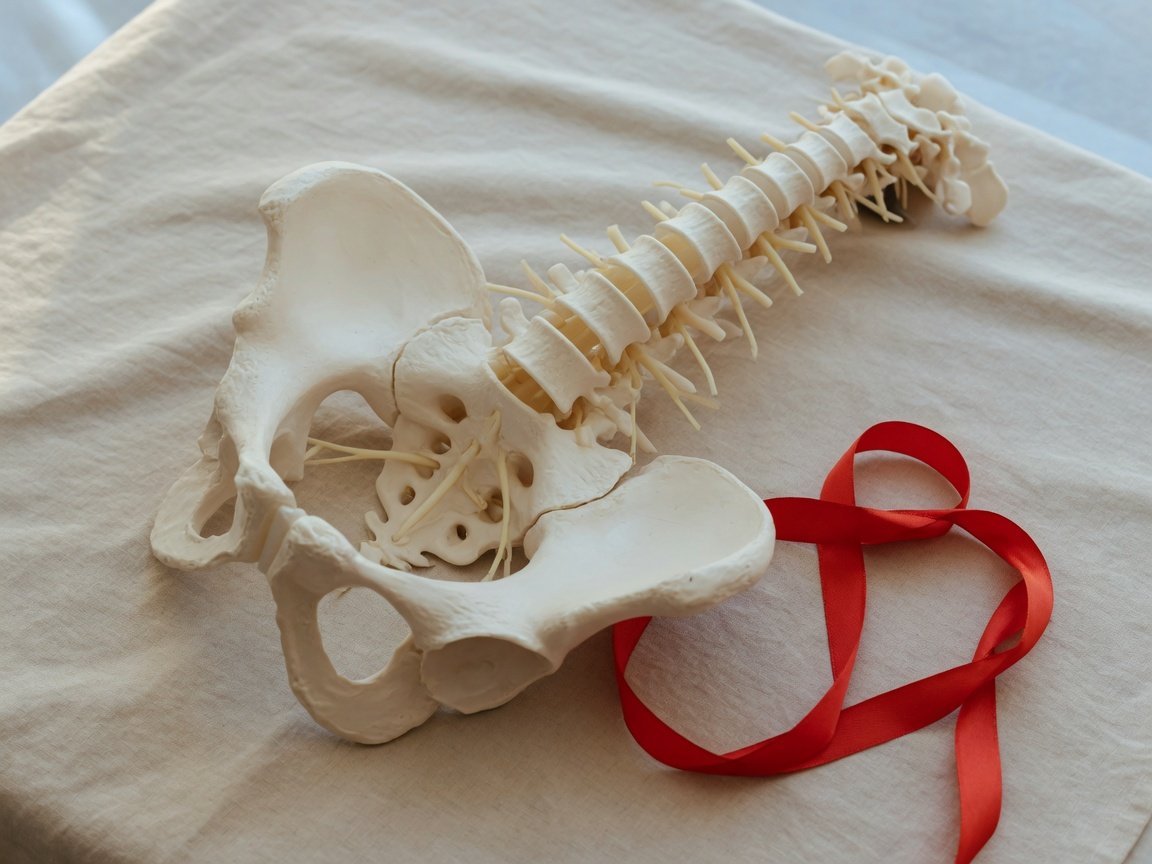
The SI joints connect the sacrum, the triangular bone at the base of the spine, to the iliac bones of the pelvis. They transfer load between the upper body and legs and normally move very little.
MRI can show both active changes and older structural changes. Common report terms include:
- Bone marrow edema: increased fluid signal within bone, sometimes described as swelling. It can occur with inflammation, mechanical stress, or injury.
- Subchondral sclerosis: denser bone just beneath the joint surface. This does not, by itself, establish inflammatory arthritis.
- Erosions: areas where the bone surface has been damaged.
- Ankylosis: fusion across a joint.
- Preserved joint spaces: the space between the bones remains maintained, without marked narrowing or fusion.
Three different explanations for SI joint changes
1. Active inflammatory sacroiliitis
Sacroiliitis means inflammation of an SI joint. It can occur in axial spondyloarthritis, a group of inflammatory rheumatic conditions affecting the spine and SI joints. Other causes, including infection, also exist, so the term is not a diagnosis of one particular disease.
On MRI, active inflammatory sacroiliitis often involves marrow edema in a characteristic location and distribution beside the joint. Erosions and other structural findings can add supporting evidence. A small spot of edema alone does not prove spondyloarthritis. Physical loading, pregnancy-related changes, and other mechanical stresses can sometimes produce edema too.
Symptoms that raise suspicion for inflammatory back pain include prolonged morning stiffness, improvement with movement rather than rest, and pain during the second half of the night. Psoriasis, inflammatory bowel disease, previous eye inflammation called uveitis, and family history can also provide clues.
2. Degenerative or mechanical changes
SI joints can develop changes from repeated loading, altered movement patterns, or aging. Reports may describe mild sclerosis, small bone spurs, joint-surface irregularity, or degenerative change. These findings are different from evidence of an immune-driven inflammatory disease.
Mechanical pain often varies with position or activity, although symptom patterns overlap. Mild imaging changes are also found in people without pain. Their presence does not prove that the SI joint is the main pain source.
3. Osteitis condensans ilii
Osteitis condensans ilii, often shortened to OCI, is a generally benign pattern of bone thickening beside the SI joint. It classically forms triangular areas of sclerosis on the iliac, or pelvic-bone, side, often on both sides, while joint spaces remain preserved.
OCI is associated with mechanical loading and is often seen after pregnancy, but it can occur in people who have never been pregnant. Despite its name, it is not the same as inflammatory arthritis. It may accompany lower-back or buttock pain, or be an incidental finding.
OCI usually lacks the destructive erosions or fusion associated with established inflammatory sacroiliitis. However, some OCI cases have nearby marrow edema, making the overall pattern important. Mild sclerosis alone is not enough to confirm OCI.
Common questions about a reassuring or unclear MRI
“No convincing active sacroiliitis”—does that rule out rheumatic disease?
It means the scan does not show a convincing active inflammatory pattern at the time of imaging. That is reassuring, particularly when joint spaces are preserved and there are no erosions or fusion.
It does not exclude every rheumatic condition or completely rule out early axial spondyloarthritis. A clinician may consider symptoms, examination, inflammatory blood tests, and sometimes HLA-B27 testing. No single test establishes or excludes the diagnosis by itself.
What if one report mentions edema and another says there is none?
Differences can reflect different scan dates, imaging protocols, image quality, or interpretation of a subtle finding. A general pelvic MRI and a dedicated SI joint MRI are not interchangeable.
The useful next step is comparison of the actual images by a qualified radiologist, not choosing the most reassuring summary. An AI-generated explanation should not override the signed report; discrepancies need professional review.
Can these findings explain pain down the leg?
SI joint pain can spread into the buttock, groin, or thigh and sometimes farther down the leg. But pain reaching the knee or ankle—especially with tingling, numbness, weakness, or a heavy sensation—also calls for consideration of other causes.
Possibilities include lumbar nerve irritation, hip conditions, peripheral nerve problems, or muscle and tendon pain. A pelvic MRI does not fully assess lumbar discs and nerve roots. Conversely, a tiny disc bulge without nerve compression may be incidental rather than the explanation.
Does a mostly normal pelvic MRI exclude hip problems or surgical adhesions?
No. A routine pelvic MRI may not answer specific questions about hip shape, small labral injuries, or postoperative adhesions. The appropriate examination and imaging depend on the suspected problem. Normal-looking muscles and bones are reassuring, but they do not exclude every source of pelvic pain.
When to talk to your doctor
Discuss persistent pain, prolonged morning stiffness, radiating symptoms, or conflicting reports with your clinician. Examination can help determine whether SI joints, the spine, hips, or another structure needs closer assessment.
Seek urgent care for new or worsening leg weakness, numbness around the groin or saddle area, loss of bladder or bowel control, fever with severe back pain, or sudden marked leg swelling.
Get AI-powered analysis of your CT or MRI scan
Upload your DICOM files and receive a clear, patient-friendly report in minutes.
Analyze my scan