Meniscus Tears, Locking, Catching, and Knee Cysts
Knee locking, catching, clicking, and small cysts can be confusing. Learn how meniscus tears and MRI wording fit together.

Have your own scan or report? Get a clear, plain-language explanation in minutes.
Why locking, catching, and clicking raise concern
Knee symptoms such as locking, catching, clicking, pain with walking, or a feeling that the knee may give way can be worrying, especially after a pivot, twist, tackle, or fall. Many people wonder whether these symptoms mean a meniscus tear, an ACL injury, a cartilage problem, or a cyst behind the knee.
This article is general education, not a diagnosis. Your own answer depends on your examination, full MRI study, prior surgery, symptoms, and your clinician’s assessment.
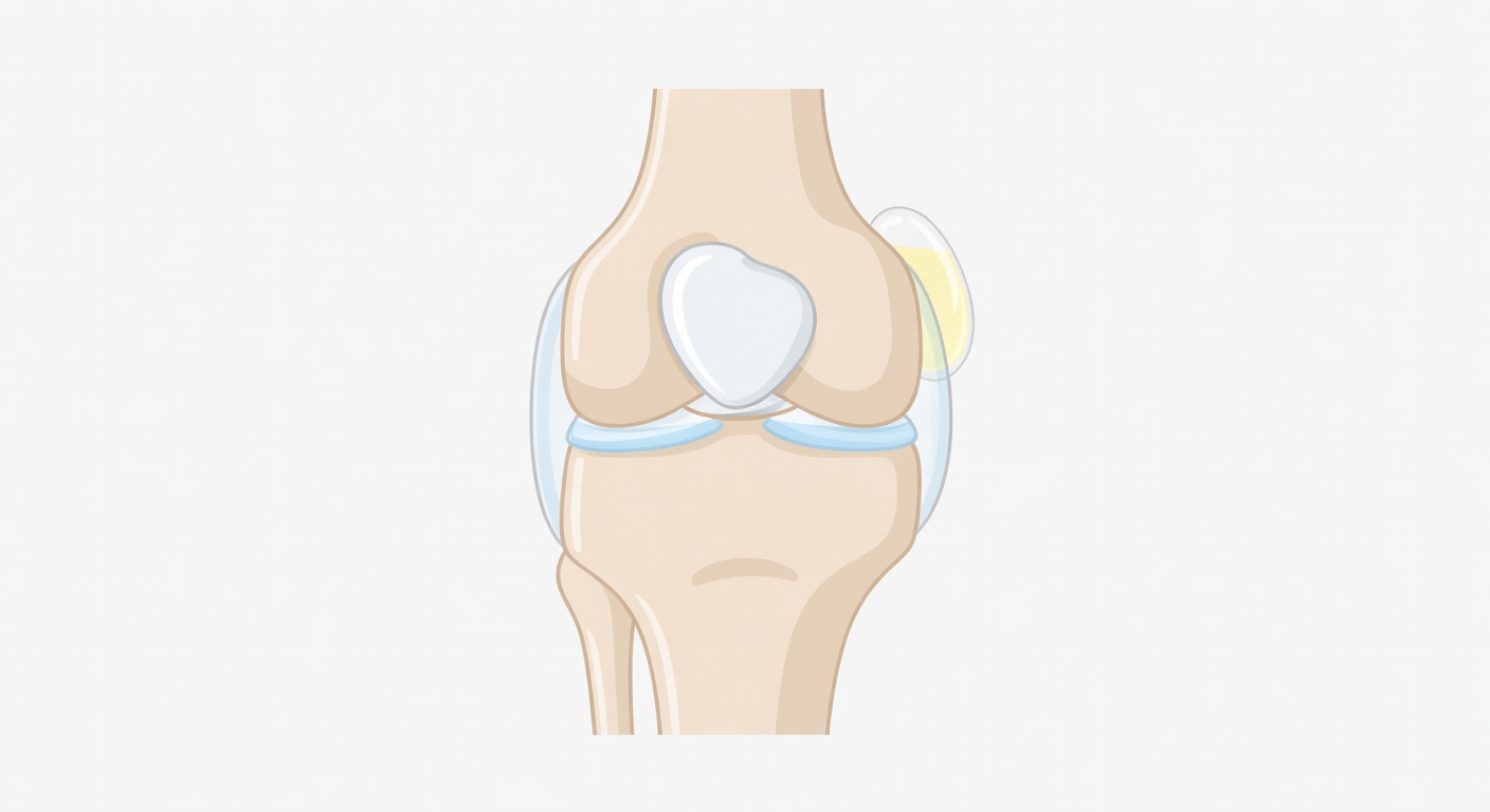
The meniscus is a C-shaped cushion of cartilage-like tissue between the thigh bone and shin bone. Each knee has a medial meniscus on the inner side and a lateral meniscus on the outer side. A meniscus tear can happen suddenly during twisting, or it can develop gradually with wear and joint changes.
Key idea: Mechanical symptoms can fit with a meniscus tear, but they are not specific. ACL injuries, cartilage defects, loose bodies, kneecap tracking problems, swelling, and irritated soft tissues can also cause catching or clicking.
Common symptoms of a meniscus tear
Meniscus tears do not all feel the same. Some are painful but stable. Others can move or fold into the joint and cause more dramatic mechanical symptoms.
Symptoms that may occur
- Joint line pain: pain along the inner or outer edge of the knee, often worse with twisting or squatting.
- Catching or clicking: a sensation that something catches during bending or straightening.
- Locking: the knee may feel stuck or unable to fully straighten or bend.
- Swelling or effusion: extra fluid inside the knee, sometimes after activity.
- Giving way: a feeling of instability, which can occur with pain, swelling, meniscus injury, or ligament injury such as an ACL tear.
- Pain with walking: especially on uneven ground, stairs, or when turning.
True locking, where the knee physically cannot move through its range, is more concerning for a displaced meniscal fragment or loose body. A feeling of catching or stiffness can also happen from swelling, inflammation, or cartilage irritation.
Could a subtle meniscus tear cause locking and catching?
Yes, a subtle meniscus tear can sometimes contribute to catching, clicking, or pain. However, symptoms alone cannot prove that a tear is present or that it is the main pain generator.
Some tears are obvious on MRI, such as a displaced bucket-handle tear, where a fragment flips into the center of the knee. Other tears are more subtle, especially horizontal, small radial, flap, or root-region tears. These may be harder to see if the tear is tiny, if there is motion during the scan, if only limited images are reviewed, or if prior surgery creates artifact.
After ACL reconstruction or other surgery, reports may mention susceptibility artifact. This means metal or surgical material can create distortion on MRI, which may limit detail in nearby structures. That does not mean the scan is useless, but it can make small findings harder to confirm.
What does an MRI report mean by no displaced fragment?
When a report says no displaced meniscal fragment, it means the radiologist did not see a large piece of meniscus flipped out of place. This is reassuring because displaced fragments are one of the classic causes of true mechanical locking.
However, this phrase does not always mean there is no meniscus tear at all. A small, nondisplaced tear may not create a visible fragment. It may still cause pain or irritation, depending on its location and the rest of the knee.
What does no macro-tear mean?
Some reports use wording such as no macro-tear, kein Makroriss, or triangular configuration of the menisci. In plain language, this usually means the menisci keep their normal shape and there is no large or obvious tear seen on the images.
A German question like schließt der Befund einen kleinen Meniskusschaden aus? means, does the report rule out a small meniscus injury? The careful answer is: it may make a large or displaced tear unlikely, but it may not completely exclude a very small or subtle meniscal injury. MRI is very helpful, but it is not perfect, and radiology reports often communicate levels of confidence rather than absolute certainty.
If a report says there is no meniscal dislocation and no macro-tear, that is generally reassuring against a major displaced tear. If symptoms strongly suggest a meniscus problem, the orthopedic clinician may still correlate the MRI with physical tests, range of motion, tenderness location, and activity-related symptoms.
Why a single MRI series may not answer the whole question
Knee MRI is usually interpreted using multiple sequences and planes: sagittal, coronal, axial, and different fluid-sensitive or T1-weighted images. Each sequence highlights different tissues.
For example, an axial series may show joint fluid and kneecap alignment well, but may be limited for meniscal tears. A sagittal ACL-focused series may show the ACL clearly, but may not fully evaluate the meniscus. Coronal fluid-sensitive images may be better for collateral ligaments and parts of the meniscus, but still need to be interpreted with the full exam.
This is why a report may say meniscal pathology cannot be excluded on this single series or subtle tear cannot be excluded without the complete MRI. That wording is not necessarily alarming. It means the provided images are incomplete for a definitive call.
How ACL injuries and meniscus tears overlap
A pivot injury followed by swelling, giving way, and mechanical symptoms raises concern for internal knee injury. ACL tears and meniscus tears can occur together. An ACL injury can cause instability, while a meniscus tear can cause pain, catching, or locking. Swelling itself can also make the knee feel tight or unreliable.
Physical exam tests such as Lachman, Apley, McMurray, and patellar compression are pieces of the puzzle. A positive test does not stand alone as a final diagnosis, but it helps guide whether the main concern is ligament instability, meniscal irritation, cartilage pain, or kneecap-related pain.
When an orthopedic surgeon recommends meniscus debridement, repair, or another procedure, that decision is usually based on the combination of symptoms, exam, imaging, activity goals, and whether the suspected tear is likely to heal or continue causing mechanical problems.
Does a small cyst-like area suggest a meniscus tear?
A small cyst-like fluid area near the knee can be confusing. It may be described as a Baker cyst, popliteal cyst, ganglion cyst, parameniscal cyst, or small fluid-signal focus. These are not all the same, but they share one feature: they contain fluid-like material.
Baker cysts and popliteal cysts
A Baker cyst, also called a popliteal cyst, is a fluid-filled swelling behind the knee. It often forms when extra joint fluid tracks backward into a small bursa. It can be associated with knee irritation from arthritis, meniscus tears, cartilage wear, inflammatory conditions, or injury. But the cyst itself does not prove a meniscus tear.
Parameniscal cysts
A parameniscal cyst is a fluid collection next to the meniscus. This type has a stronger association with a meniscal tear because fluid can pass through a tear and collect beside the meniscus. Even then, the MRI and symptoms need to match.
Ganglion-type fluid
Ganglion cysts are benign fluid-filled areas that can occur around joints and tendons. A small posterior or posteromedial ganglion-type focus may be an incidental finding, meaning it is present but not necessarily the cause of symptoms.
So, to answer the common question: a small cyst-like area can be associated with a meniscus tear, but it does not automatically prove one. Its location matters. A cyst directly along the meniscal margin is more suspicious for a meniscal connection than a small nonspecific cyst behind the knee.
What if the pain feels exactly like a meniscus tear?
Many people say their pain feels like a meniscus tear. That description is important and should be taken seriously, but several knee problems can mimic meniscus pain. Cartilage wear in the medial compartment, bone stress reaction, MCL sprain, kneecap cartilage irritation, joint swelling, and ACL-related instability can all cause pain with walking, twisting, or stairs.
Radiology reports may also mention chondrosis, chondral thinning, osteophytes, marrow edema-like signal, synovitis, or effusion. These findings can contribute to pain and swelling, even when no displaced meniscus fragment is seen.
Putting the report and symptoms together
The most useful approach is to combine three sources of information:
- Your story: how the injury happened, whether there was a pop, swelling, locking, or giving way.
- The exam: stability testing, joint line tenderness, range of motion, and kneecap tracking.
- The full MRI: not just one image or sequence, but the complete study interpreted in context.
A report that says no fracture, no displaced fragment, no large Baker cyst, and no macro-tear is often reassuring. But if locking, catching, or giving way persists, it is reasonable for the treating clinician to look closely for subtle meniscal, ligament, cartilage, or kneecap-stabilizing injuries.
When to talk to your doctor
Talk to a sports medicine clinician, orthopedic specialist, or your regular doctor if you have persistent locking, catching, swelling, giving way, or pain that limits walking or normal activity. Seek prompt medical care if the knee is truly stuck, you cannot bear weight, swelling is severe, there is fever or redness, or symptoms are worsening after injury.
This information is for general education and does not diagnose your knee. Your clinician can explain how your MRI report, exam findings, and treatment options fit your specific situation.
Get AI-powered analysis of your CT or MRI scan
Upload your DICOM files and receive a clear, patient-friendly report in minutes.
Analyze my scan