Ankle MRI: Peroneal Tendinosis and Tenosynovitis
Learn what peroneal tendinosis, tenosynovitis, and partial tearing mean on an ankle MRI report.

Have your own scan or report? Get a clear, plain-language explanation in minutes.
What an ankle MRI can show about tendon pain
An ankle MRI report can feel confusing when it says the ligaments are intact, but also describes tendon irritation, fluid, or a partial tear. Many people read this and wonder: if nothing is broken and the ligaments look normal, why does walking still hurt?
This article explains common MRI terms such as peroneal tendinosis, tenosynovitis, and partial tear in plain language. It is general education, not a diagnosis, and it cannot replace a clinician who can examine your ankle and match the MRI findings to your symptoms.
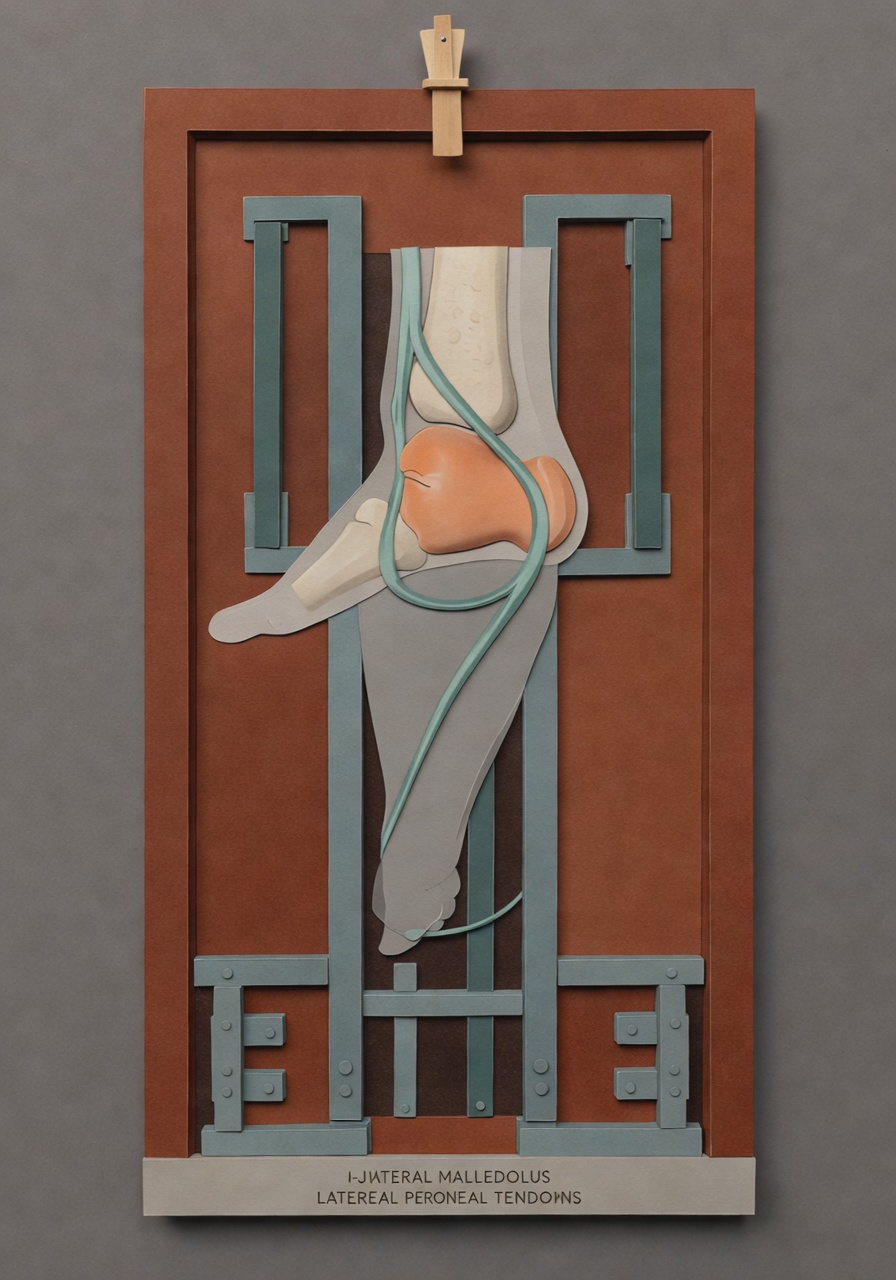
Meet the peroneal tendons: peroneus brevis and longus
The peroneal tendons run along the outside and back of the ankle, behind the bony bump on the outer side called the lateral malleolus. There are two main tendons:
- Peroneus brevis: This tendon helps turn the foot outward and supports the outside of the foot. It attaches near the base of the fifth metatarsal, the long bone on the outer side of the foot.
- Peroneus longus: This tendon also helps turn the foot outward, then travels under the foot to help support the arch and stabilize the first ray near the big toe side.
These tendons are important for balance, walking on uneven ground, pushing off, and preventing the ankle from rolling inward. Because they pass through a tight tunnel behind the outer ankle bone, they can become irritated after sprains, overuse, high-impact activity, foot shape issues, or prior injury.
What does peroneal tendinosis mean?
Tendinosis means the tendon shows wear-and-tear type change or degeneration. On MRI, the tendon may look thickened, irregular, or show abnormal signal inside it. This does not always mean a sudden injury happened. It often suggests the tendon has been under stress for a while.
Tendinosis can be painful, but MRI appearance and pain do not always match perfectly. Some people have mild-looking tendinosis with significant symptoms, while others have more obvious imaging changes but less pain. That is why the location of your pain, your activity level, and your physical exam matter.
What is tenosynovitis?
Tenosynovitis means inflammation or irritation of the tendon sheath, the thin lining around certain tendons. On MRI, it often appears as fluid around the tendon. A report may say mild tenosynovitis, marked tenosynovitis, or fluid around the peroneal tendons.
In simple terms: tendinosis is change within the tendon, while tenosynovitis is irritation around the tendon.
Tenosynovitis can cause aching, burning, swelling, or a feeling of fullness along the outside of the ankle. It may be more noticeable with walking, stairs, uneven ground, or activity that requires side-to-side ankle control.
What does a partial tear mean?
A partial tear means some tendon fibers are damaged, but the tendon is not completely torn through. This is different from a full-thickness rupture, where the tendon is completely disrupted.
Peroneus brevis tears can sometimes be described as a split tear because the tendon may split lengthwise as it travels behind the outer ankle bone. A partial tear may occur along with tendinosis and tenosynovitis, because a worn or irritated tendon is more vulnerable to fiber damage.
The word partial is important. It means the tendon still has continuity. Treatment decisions depend on many factors, including pain level, ankle stability, function, activity goals, and whether symptoms improve with non-surgical care.
Why can ligaments be intact but the ankle still hurts?
An ankle MRI often has separate sections for ligaments, tendons, bones, joints, and other soft tissues. A report may say the anterior talofibular ligament, posterior talofibular ligament, tibiofibular ligaments, and deltoid ligament complex are intact. That is reassuring because it means the major stabilizing ligaments do not show a sprain or tear on the MRI.
However, intact ligaments do not rule out tendon pain. Ligaments and tendons do different jobs:
- Ligaments connect bone to bone and help stabilize joints.
- Tendons connect muscle to bone and help move and support the foot and ankle.
So a person can have normal-appearing ligaments but still have significant pain from peroneal tendinosis, tenosynovitis, a partial tendon tear, joint irritation, or other soft-tissue causes.
What about mild joint fluid or tibiotalar effusion?
The tibiotalar joint is the main ankle joint between the shin bone and the talus bone. A joint effusion means extra fluid in the joint. MRI reports may describe this as minimal, mild, small, or moderate.
A small amount of fluid can be nonspecific. It may be seen with irritation, recent activity, arthritis, inflammation, or after injury. By itself, mild fluid usually does not explain everything. It becomes more meaningful when it matches the exam, pain location, swelling, and other MRI findings.
Other findings that may appear on the same report
Mild Achilles tendinosis
The Achilles tendon runs at the back of the ankle and attaches to the heel bone. Mild Achilles tendinosis means early or low-grade tendon degeneration. It may or may not be related to outer ankle pain. If your pain is mostly at the back of the heel or calf area, it may be more relevant.
Posterior tibial tenosynovitis
The posterior tibial tendon runs along the inside of the ankle and helps support the arch. Minimal posterior tibial tenosynovitis means a small amount of fluid or irritation around that tendon. Like peroneal tenosynovitis, its importance depends on where your symptoms are.
Midfoot spurring
Spurring means small bony overgrowths, often related to chronic stress or arthritis-type change. Minimal midfoot spurring may be an incidental finding, especially if pain is not located in the midfoot.
Post-surgical hardware changes
If you have had prior ankle surgery, imaging may mention metal hardware, chronic remodeling, or posttraumatic changes. Hardware can be intact while nearby tendons or joints are still irritated. Metal can also create artifact on CT or MRI, which may limit how clearly nearby tissues are seen.
Why symptoms matter as much as the MRI words
MRI is very helpful, but it is only one part of the picture. A clinician will usually compare the report with questions such as:
- Where exactly is the pain: outside ankle, inside ankle, back of heel, or midfoot?
- Is there swelling, clicking, snapping, or a feeling of instability?
- Does pain worsen with walking, running, stairs, or uneven ground?
- Was there a recent ankle sprain or older injury?
- Have symptoms improved, stayed the same, or worsened over time?
For example, peroneal tendon findings are more likely to matter if your pain is along the outer back part of the ankle, especially if it flares with walking or side-to-side movement. If your pain is in a different area, the same MRI finding may be less central.
Common next-step discussions
Your clinician may discuss options based on the severity of symptoms and the MRI findings. These may include activity changes, bracing or support, physical therapy focused on strength and balance, footwear changes, anti-inflammatory strategies when appropriate, or referral to a foot and ankle specialist. In some cases, especially with persistent pain, mechanical symptoms, or a significant tear, a surgical opinion may be considered.
The key point is that an MRI phrase such as marked tenosynovitis or partial tear does not automatically mean one specific treatment. The best next step depends on the whole clinical picture.
How to read your report before your appointment
Before meeting with your clinician, it can help to write down:
- The exact location of pain and whether it matches the peroneal tendon area.
- What activities trigger symptoms, such as walking, stairs, sports, or uneven ground.
- Whether you feel snapping, giving way, swelling, or weakness.
- Any history of ankle sprain, fracture, surgery, or hardware.
- What has already helped or made symptoms worse.
This makes the conversation more practical and helps your clinician connect the MRI findings to your day-to-day function.
When to talk to your doctor
Talk with your doctor or a qualified health professional if ankle pain persists, limits walking, is associated with swelling or instability, or follows an injury. Seek urgent care if you cannot bear weight, have severe worsening pain, new numbness, signs of infection, or a sudden change in foot color or temperature. This information is for general education only and is not a diagnosis or personalized medical advice.
Get AI-powered analysis of your CT or MRI scan
Upload your DICOM files and receive a clear, patient-friendly report in minutes.
Analyze my scan