Explicación de la RM del LCA, el menisco y la luxación rotuliana
Guía en lenguaje sencillo sobre términos de la RM de rodilla después de lesiones por torsión, incluidas las roturas del LCA, las roturas de menisco, los edemas óseos, el derrame articular y la luxación rotuliana.

Comprender una RM de rodilla después de una lesión por torsión
Un informe de RM de rodilla puede resultar abrumador, especialmente después de una lesión deportiva o una torsión repentina. Términos como rotura del LCA, rotura del menisco lateral, contusión ósea, esguince del LCM, derrame articular grande o patrón de luxación-reducción rotuliana pueden aparecer en el mismo informe. Para adultos activos, deportistas adolescentes y padres, ayuda comprender qué suelen significar estas palabras y por qué los médicos evalúan la RM completa, no solo una imagen o una frase.
Este artículo es información educativa general, no un diagnóstico. Su propia RM debe ser interpretada por un radiólogo y correlacionarse con cómo ocurrió la lesión, la exploración física, los síntomas y los objetivos.
Por qué los informes de RM de rodilla usan tantos términos
La rodilla tiene varias estructuras importantes concentradas en un espacio pequeño: ligamentos, meniscos, cartílago, hueso, tendones y el sistema de seguimiento de la rótula. Una lesión por torsión puede afectar a más de una estructura al mismo tiempo. La RM es útil porque puede mostrar tejidos blandos y cambios en la médula ósea que quizá no aparezcan en las radiografías convencionales.
Sin embargo, los hallazgos de la RM no siempre son blanco o negro. Un informe puede decir sospechoso de, no se puede excluir o evaluación incompleta. Esto no significa que el informe sea deficiente. A menudo significa que el radiólogo está siendo cuidadoso, especialmente si el movimiento, la hinchazón o las secuencias de imagen limitadas dificultan la valoración de una estructura.
Idea clave: Una RM de rodilla es una parte del rompecabezas. La exploración física a menudo ayuda a determinar si un ligamento es realmente inestable o si una rotura de menisco está causando síntomas mecánicos.
Qué significa alteración interna
Alteración interna es una expresión médica amplia. Significa que algo dentro de la articulación de la rodilla puede estar lesionado o alterado. No es un diagnóstico único. Puede incluir lesiones como una rotura del LCA, una rotura de menisco, una lesión del cartílago, un cuerpo libre o un esguince ligamentoso.
Si un informe dice que no se puede excluir una alteración interna, generalmente significa que las imágenes o el patrón de lesión generan preocupación, pero la estructura exacta requiere una revisión más completa o correlación clínica. En muchos estudios de RM de rodilla, los radiólogos revisan imágenes sagitales, coronales y axiales para comprender el patrón completo.
Lesión del LCA en la RM
El ligamento cruzado anterior, o LCA, ayuda a controlar el movimiento hacia delante y la rotación de la tibia bajo el fémur. Las lesiones del LCA ocurren con frecuencia al pivotar, aterrizar, cambiar de dirección o desacelerar de forma repentina.
En la RM, una lesión del LCA puede describirse como:
- Esguince: El ligamento se ve hinchado o irritado, pero aún puede tener algunas fibras intactas.
- Rotura parcial: Algunas fibras están rotas, mientras que otras permanecen unidas.
- Rotura completa o ruptura: La banda normal, tensa y oscura está interrumpida o no se ve claramente.
- LCA mal definido o heterogéneo: El ligamento no se ve normal, pero la gradación puede requerir una revisión completa de la RM.
Las lesiones del LCA suelen acompañarse de otros signos, como líquido articular y contusiones óseas en áreas características. También pueden producirse lesiones de menisco o cartílago, por lo que es importante contar con un informe completo.
Roturas de menisco y quistes parameniscales
Los meniscos son amortiguadores en forma de C entre el fémur y la tibia. Hay un menisco medial en el lado interno y un menisco lateral en el lado externo. Una lesión por torsión puede comprimir o desgarrar el menisco.
Los informes de RM pueden describir una rotura de menisco como horizontal, vertical, radial, compleja, desplazada o que afecta al cuerno anterior, el cuerpo o el cuerno posterior. Estas palabras describen la forma y la ubicación de la rotura. Una rotura del menisco lateral significa que está afectado el menisco externo.
A veces, un informe menciona un quiste parameniscal. Se trata de una pequeña bolsa llena de líquido junto al menisco. Puede ocurrir cuando el líquido articular se desplaza a través de una rotura meniscal. El quiste en sí suele ser una pista de que el menisco cercano debe evaluarse cuidadosamente.
Los síntomas no siempre coinciden perfectamente con los hallazgos de la RM. Algunas roturas causan chasquidos, bloqueo, hinchazón o dolor con la torsión. Otras pueden ser menos sintomáticas. Las decisiones de tratamiento dependen del patrón de la rotura, los síntomas, la edad, el nivel de actividad y las lesiones asociadas.
Contusiones óseas: ¿qué es un edema óseo?
Una contusión ósea, a menudo llamada edema óseo, significa que la RM muestra hinchazón o lesión dentro de la médula ósea. No es lo mismo que una fractura grande desplazada, pero aun así puede ser dolorosa y significativa.
Las contusiones óseas pueden ayudar a contar la historia de cómo ocurrió la lesión. Por ejemplo:
- La contusión en el cóndilo femoral lateral y la tibia cercana puede verse en lesiones por pivote, incluidos los patrones de lesión del LCA.
- La contusión alrededor de la rótula y la parte anteroexterna del fémur puede sugerir una luxación reciente de la rótula que volvió a su lugar.
- La contusión cerca de la meseta tibial puede reflejar fuerzas de impacto, torsión o compresión.
Los radiólogos también buscan cuidadosamente una lesión osteocondral, que significa daño que afecta al cartílago y al hueso subyacente. A veces, un pequeño fragmento de cartílago y hueso puede convertirse en un cuerpo libre dentro de la articulación. Esta es una de las razones por las que una gran hinchazón después de un episodio de inestabilidad rotuliana se toma en serio.
Derrame articular grande: por qué importa el exceso de líquido
Un derrame de rodilla significa que hay líquido adicional dentro de la articulación. Después de un traumatismo, un derrame moderado o grande puede ser una señal de que la rodilla ha sufrido una lesión o irritación interna significativa. El líquido puede acumularse en el receso suprarrotuliano, el espacio por encima de la rótula, por lo que los informes suelen mencionar esa zona.
El líquido por sí solo no identifica la lesión exacta. Puede aparecer con roturas de ligamentos, roturas de menisco, lesiones de cartílago, luxaciones rotulianas, contusiones óseas, inflamación y otras causas. Pero cuando aparece un derrame grande después de una lesión deportiva repentina, los clínicos suelen prestar mucha atención al LCA, los meniscos, el cartílago y los estabilizadores rotulianos.
Esguince del LCM en la RM
El ligamento colateral medial, o LCM, recorre el lado interno de la rodilla. Ayuda a resistir las fuerzas que empujan la rodilla hacia dentro. Un esguince del LCM puede aparecer en la RM como hinchazón alrededor del ligamento, engrosamiento o lesión parcial de las fibras.
Los informes pueden decir que las fibras del LCM son continuas, lo que sugiere que el ligamento no está completamente roto, incluso si hay edema circundante. El grado del esguince depende de los hallazgos de imagen y de la exploración física. Las lesiones del LCM también pueden ocurrir junto con lesiones del LCA o del menisco, especialmente después de un mecanismo de torsión o contacto.
Luxación rotuliana y patrón de reducción
Una luxación rotuliana significa que la rótula se salió de su surco normal, con mayor frecuencia hacia el lado externo de la rodilla. En muchos casos, vuelve a su lugar antes de la imagen. Por eso los informes de RM pueden usar la frase luxación rotuliana lateral transitoria o lesión por luxación-reducción.
Aunque la rótula esté de nuevo en su sitio durante la RM, la lesión puede dejar pistas:
- Contusión ósea en el lado interno de la rótula y en la parte externa del fémur.
- Hinchazón o desgarro del ligamento patelofemoral medial, también llamado LPFM.
- Derrame articular grande.
- Posible lesión del cartílago o fragmento libre.
- Hinchazón de tejidos blandos alrededor de la parte anterior e interna de la rodilla.
El LPFM es una estructura de contención de tejido blando importante que ayuda a evitar que la rótula se desplace hacia fuera. Si está esguinzado o roto, el clínico tratante puede considerar la estabilidad, los episodios repetidos, la lesión del cartílago, la alineación y las demandas de actividad al hablar sobre el tratamiento.
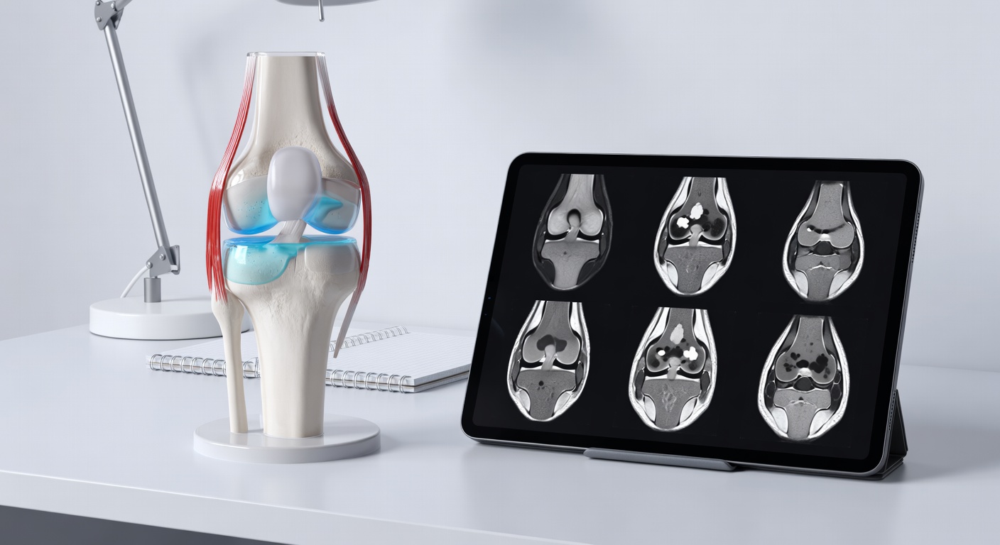
Por qué una sola serie de RM puede no ser suficiente
Los estudios de RM de rodilla suelen estar compuestos por múltiples conjuntos de imágenes, llamados secuencias, obtenidos en diferentes planos. Una serie sagital puede mostrar bien el LCA, una serie coronal puede ayudar con los ligamentos colaterales y los meniscos, y una serie axial es especialmente útil para la rótula y los estabilizadores rotulianos. Las secuencias sensibles al líquido resaltan la hinchazón y las contusiones óseas.
Si solo hay una secuencia disponible, pueden pasarse por alto lesiones importantes o sobreinterpretarse. Por eso los informes a menudo recomiendan revisar el estudio completo de RM. El informe radiológico oficial debe considerar todas las imágenes disponibles en conjunto.
Preguntas que pueden hacer los pacientes y los padres
- ¿La RM completa confirma una rotura del LCA, o se trata de un esguince o una lesión parcial?
- ¿Hay una rotura de menisco y, si es así, dónde se localiza?
- ¿Hay contusiones óseas y sugieren un patrón específico de lesión?
- ¿Hay daño del cartílago o un fragmento libre en la articulación?
- ¿El patrón sugiere una luxación de la rótula que se redujo?
- ¿Están intactos el LCM, el LPFM y otros ligamentos estabilizadores?
- ¿Qué síntomas harían más urgente el seguimiento?
Las decisiones sobre el regreso al deporte deben individualizarse y tomarse con un clínico cualificado. Los hallazgos de la RM, la fuerza, la hinchazón, la movilidad, la estabilidad, el dolor, las demandas del deporte y el progreso de la rehabilitación son todos importantes.
Cuándo hablar con su médico
Hable con un médico, un profesional de medicina deportiva o un especialista en ortopedia si presenta hinchazón persistente, inestabilidad, bloqueo, chasquidos, incapacidad para apoyar peso, empeoramiento del dolor, entumecimiento, fiebre o preocupación después de una lesión de rodilla. Busque atención médica de inmediato si tiene síntomas intensos o hinchazón que aumenta rápidamente. Este artículo es solo para educación general y no constituye un diagnóstico ni sustituye su propia evaluación médica.
Get AI-powered analysis of your CT or MRI scan
Upload your DICOM files and receive a clear, patient-friendly report in minutes.
Analyze my scan